|
|
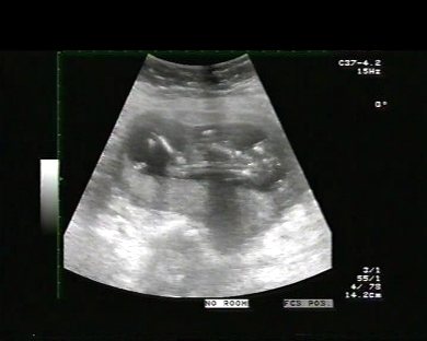
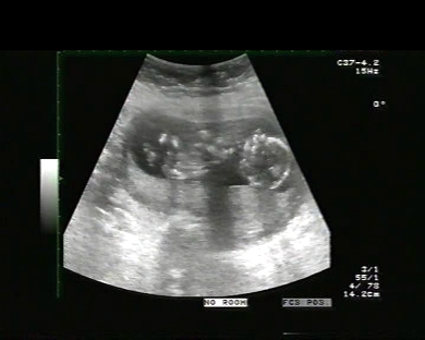
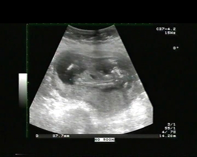
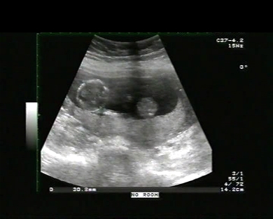
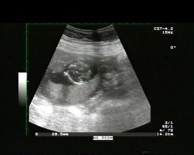
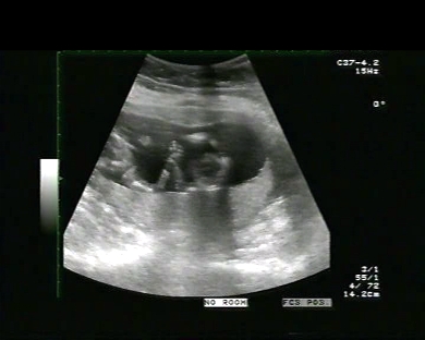
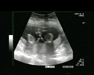
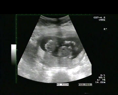
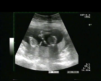
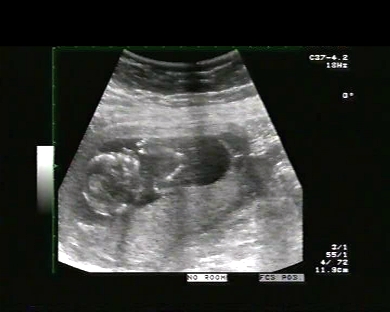
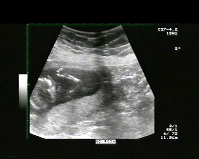
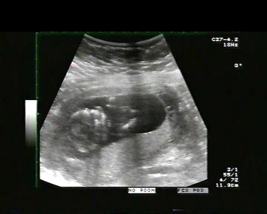
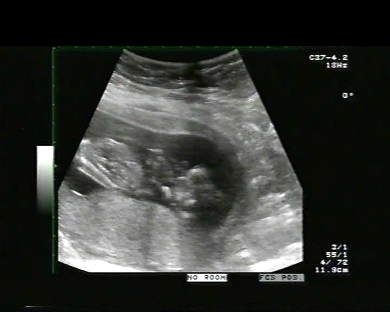
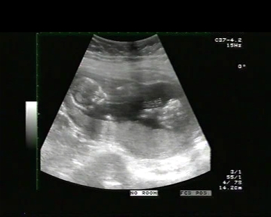
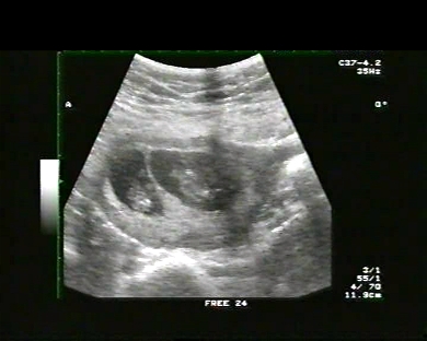
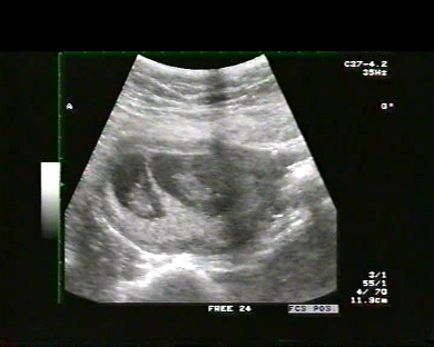
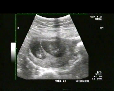
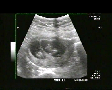
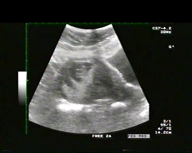
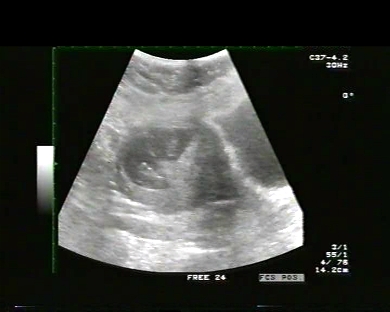
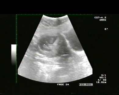
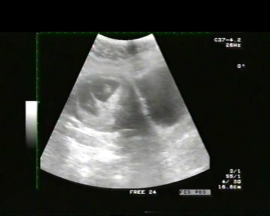
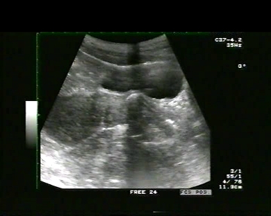
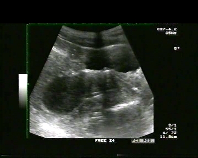
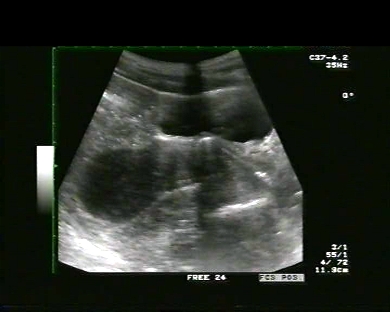
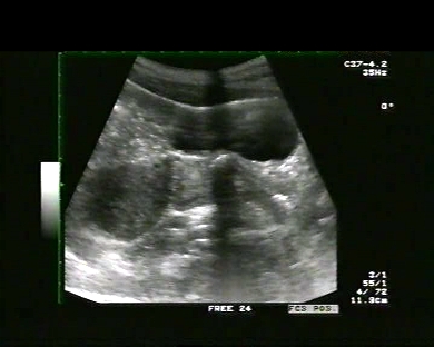
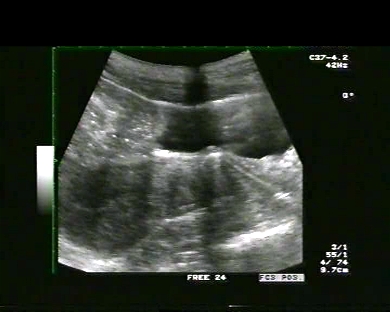
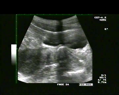
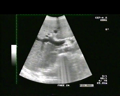
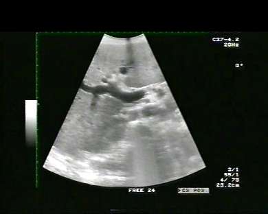
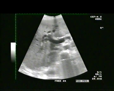
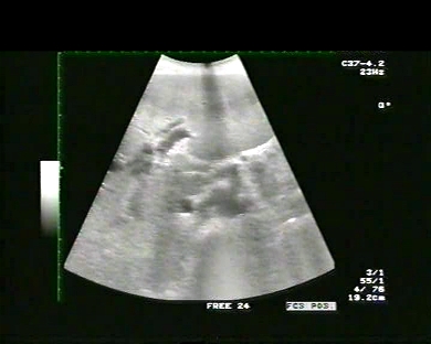
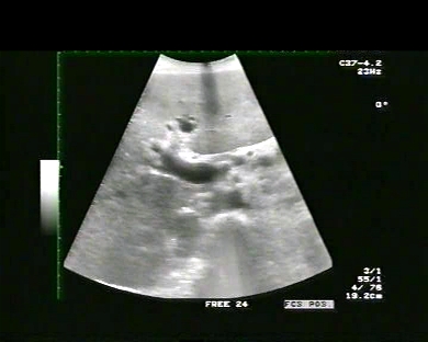
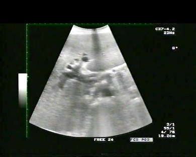
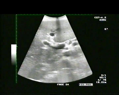
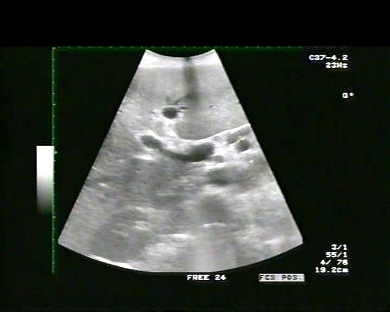
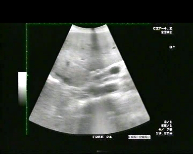
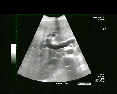
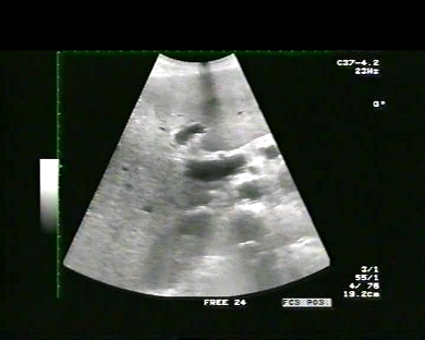
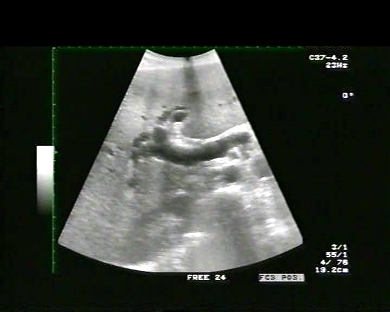
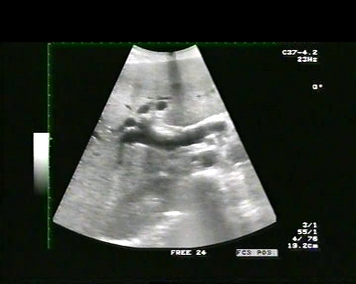
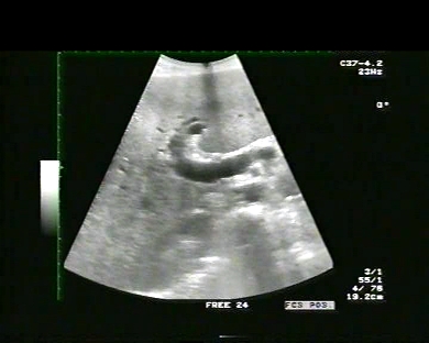
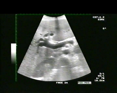
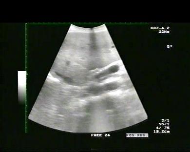
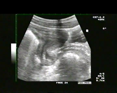
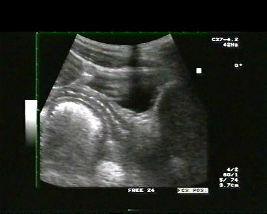
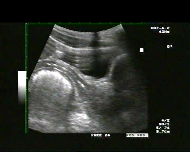
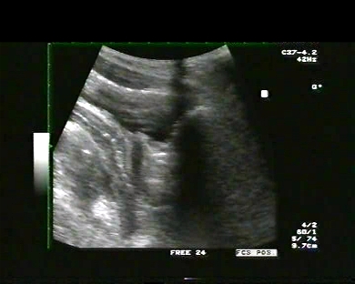
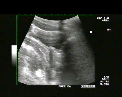
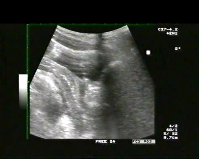
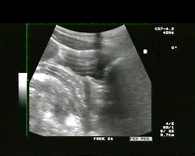
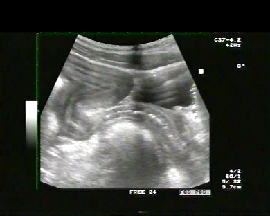
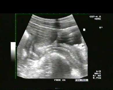
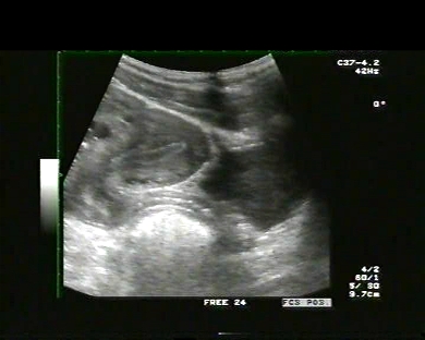
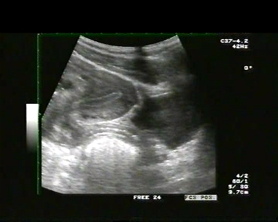
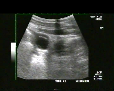
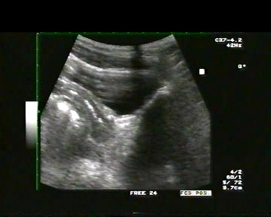
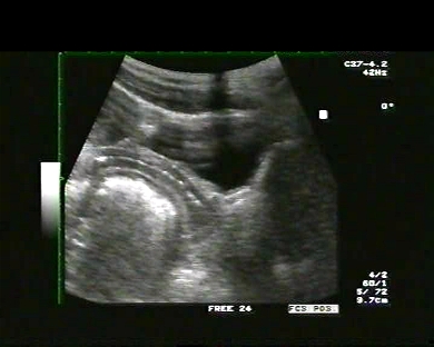
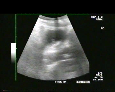
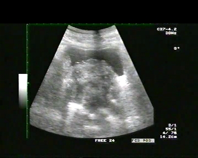
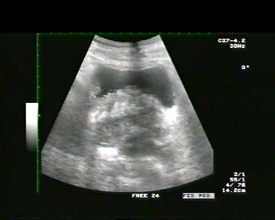
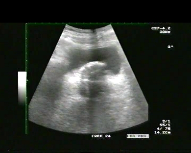
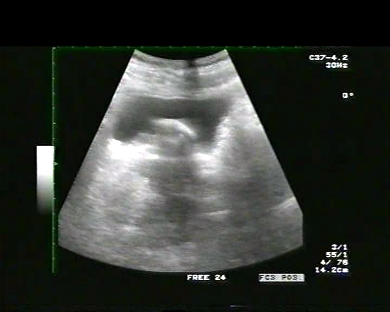
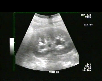
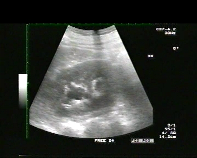
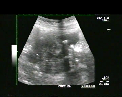
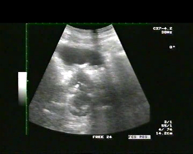
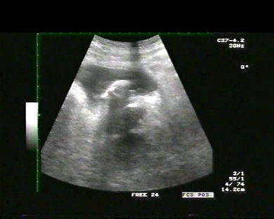
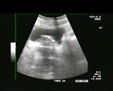
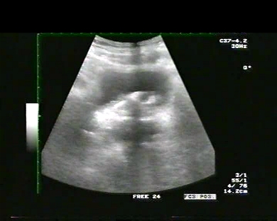
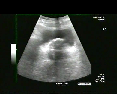
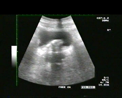
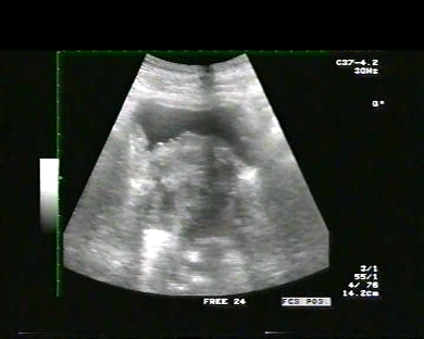
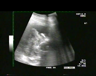
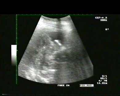
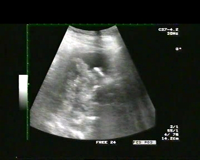
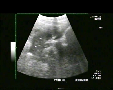
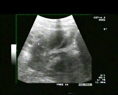
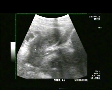
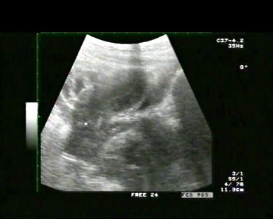
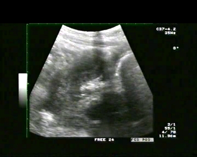
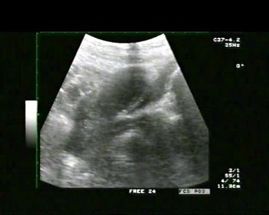
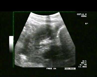
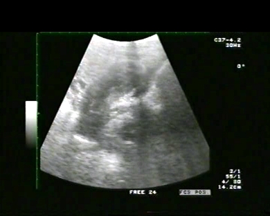
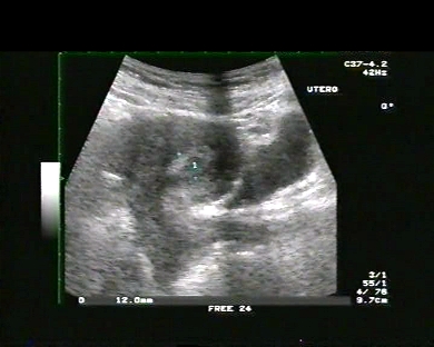
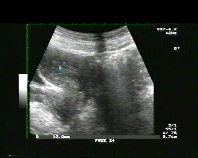
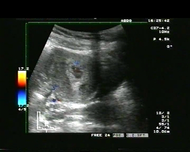
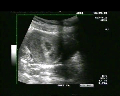
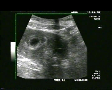
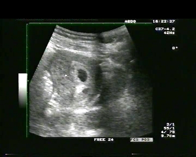
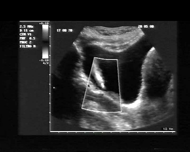
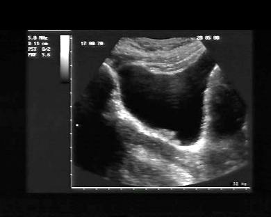
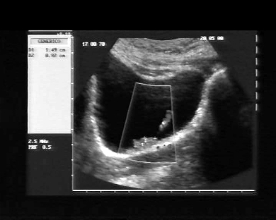
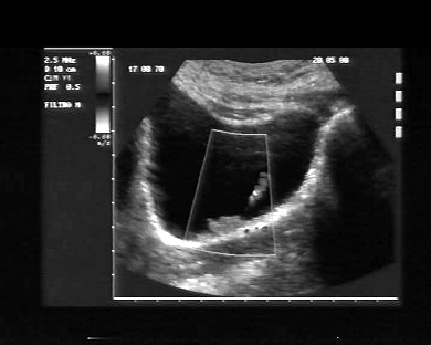
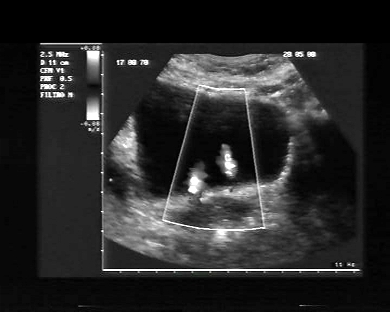
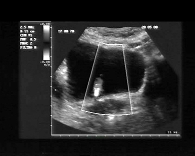
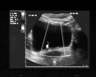
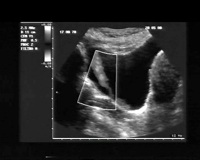
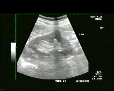
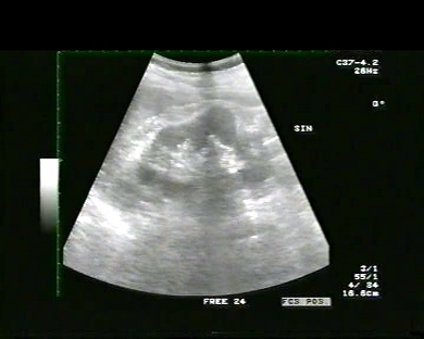
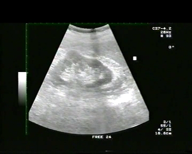
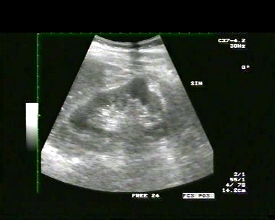
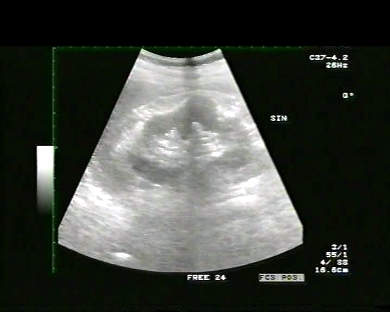
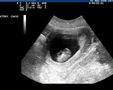
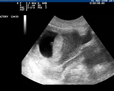
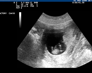
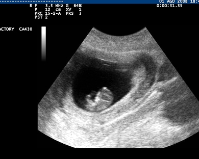
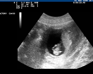
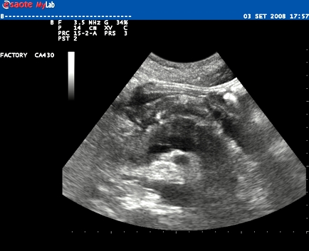
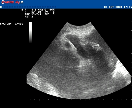
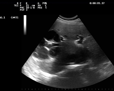
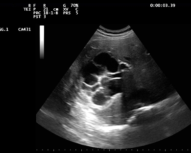
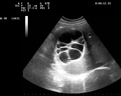
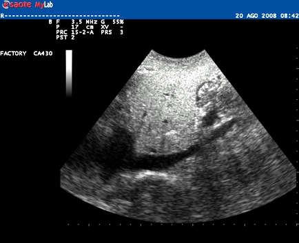
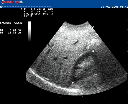
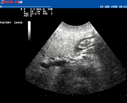
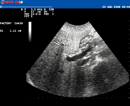
Inclusion date: 29 /12/2008Ultrasound made on: 10/11/2008 Instrument: Toshiba 380A Patient's age: F 30 years old The ultrasound examination in suprapubic position is carried out on a woman on her 14th week of pregnancy. The images and the video show a twin pregnancy with the two foetuses in the uterus. They have regular dimensions in accordance with the pregnancy stage. Presentation: Dr. Massimo Dolciotti - Ancona Digital processing: Andrea Dini - Ancona
|
|
|
|
|
|
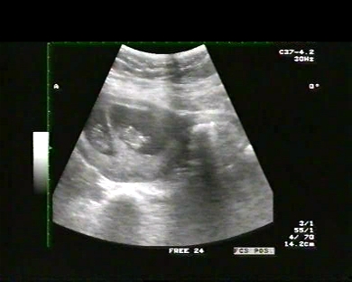
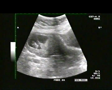
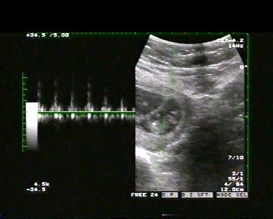
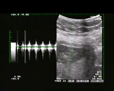
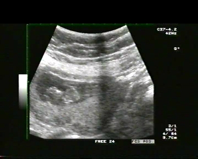
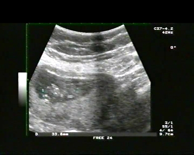
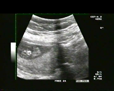
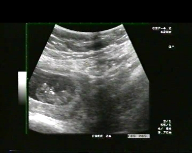
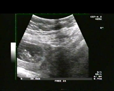
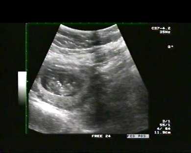
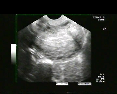
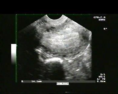
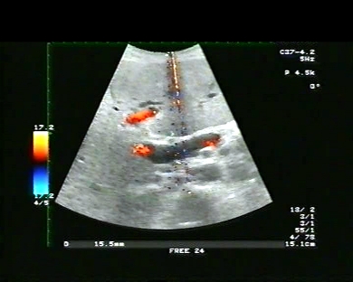
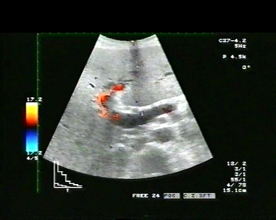
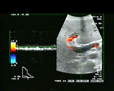
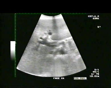
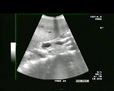
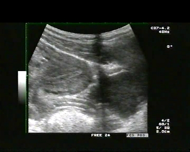
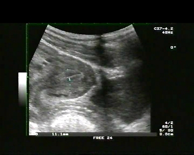
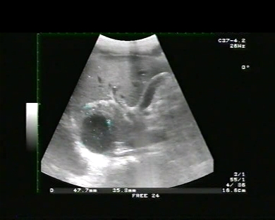
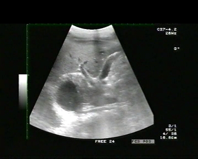
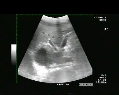
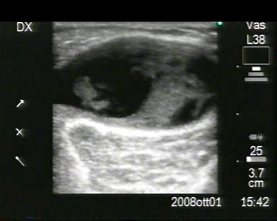
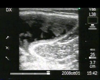
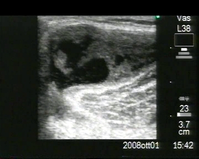
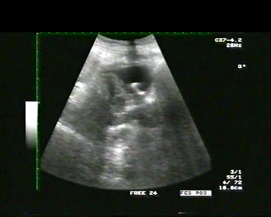
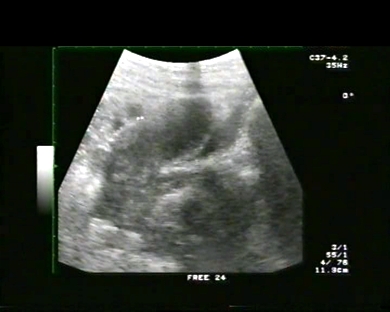
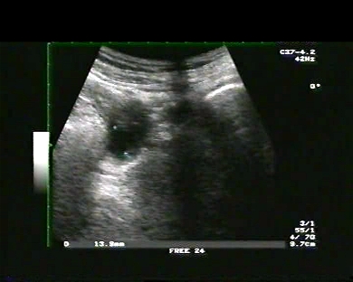
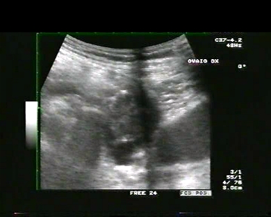
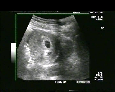
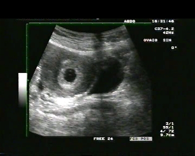
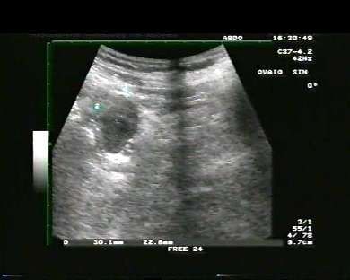
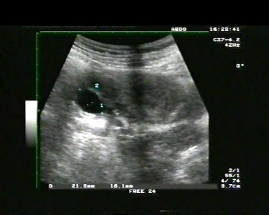
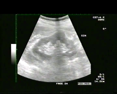
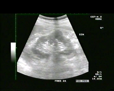
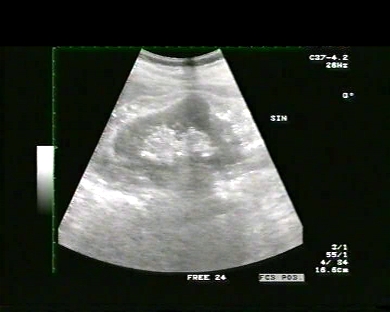
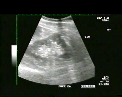
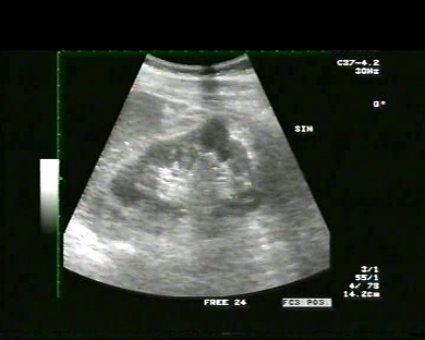
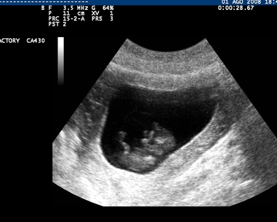
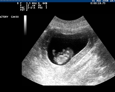
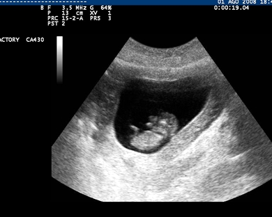
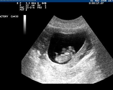
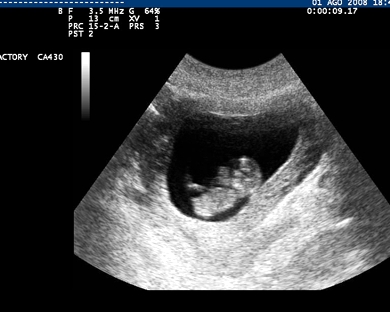
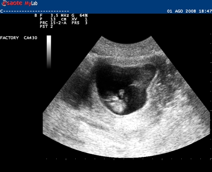
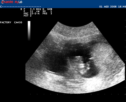
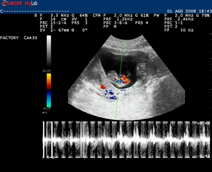
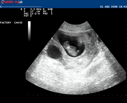
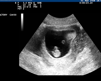
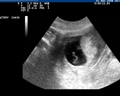
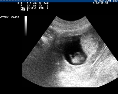
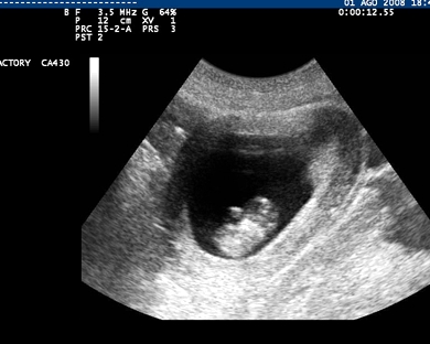
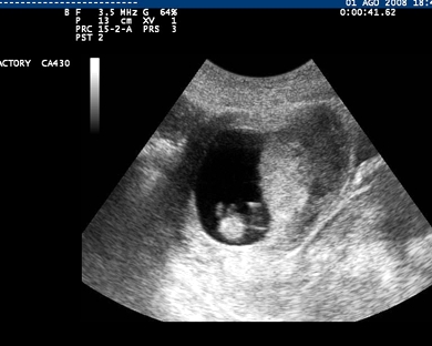
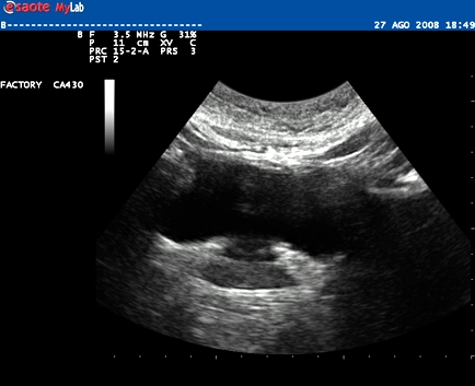
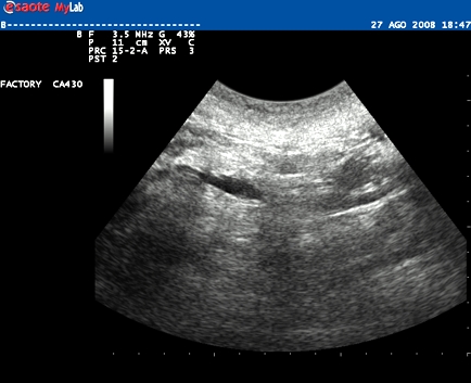
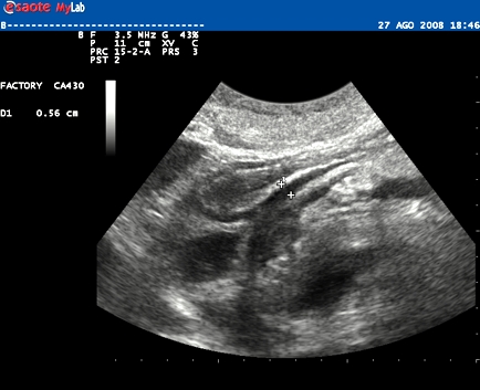
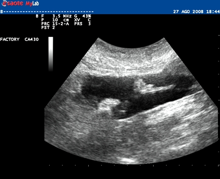
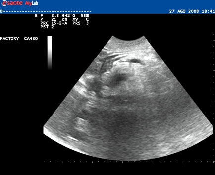
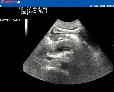
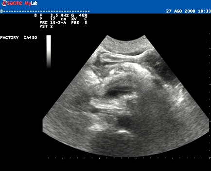
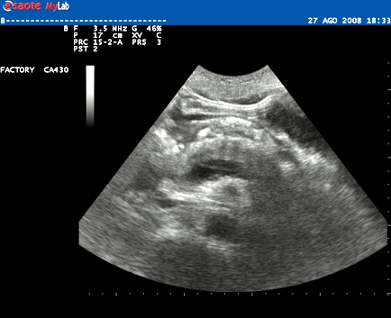
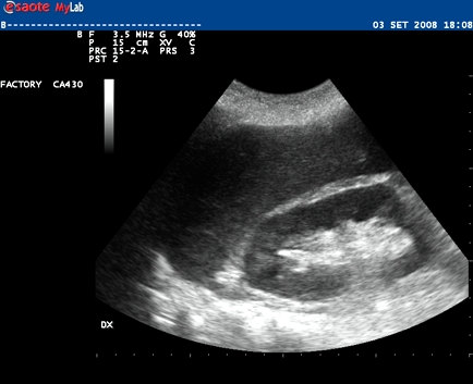
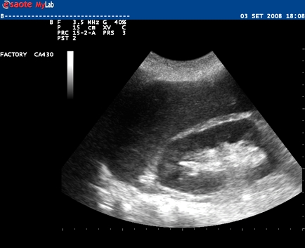
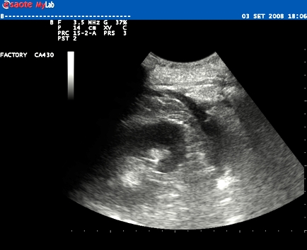
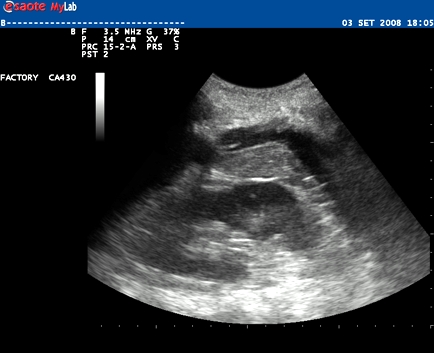
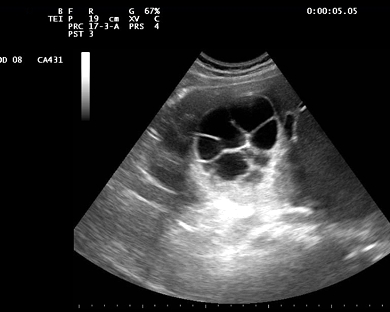
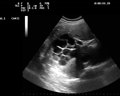
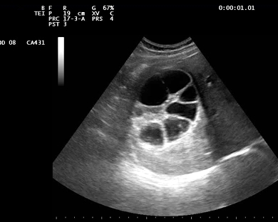
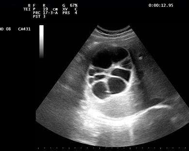
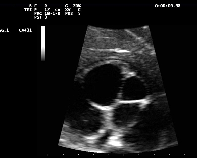
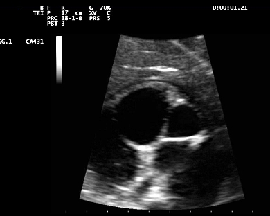
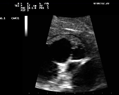
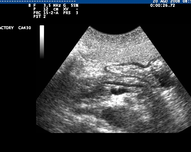
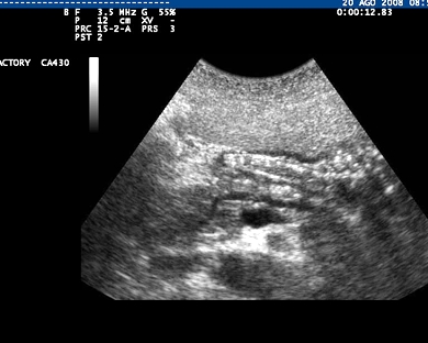
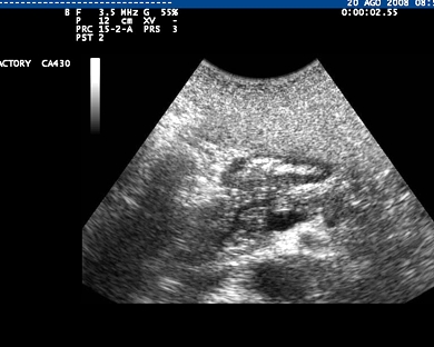
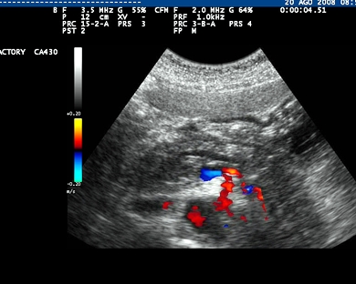
Inclusion date: 19 /12/2008Ultrasound made on: 27/09/2008 Instrument: Toshiba 380A Patient's age: F 34 years old The pelvic ultrasound examination is carried out to prove the twin pregnancy. The images and the video show the uterus with foetuses having a regular heart beat and a diameter Crown-rump length of 33mm and 38 mm respectively. Presentation: Dr. Massimo Dolciotti - Ancona Digital processing: Andrea Dini - Ancona
|
|
|
|
|
|
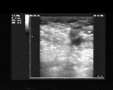
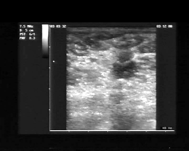
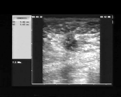
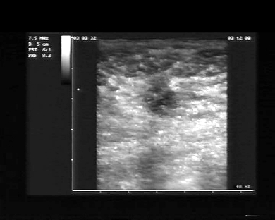
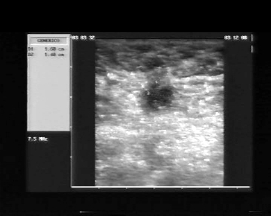
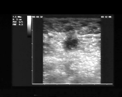
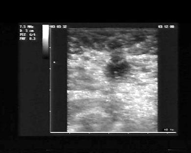
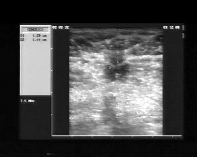
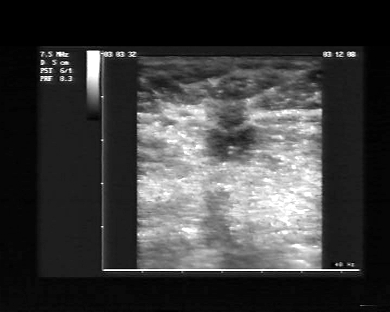
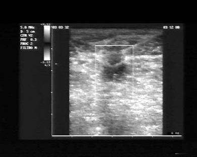
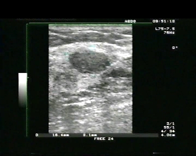
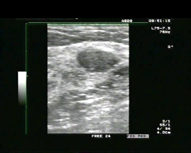
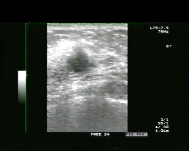
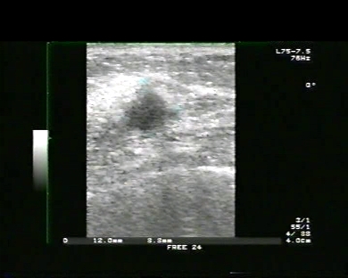
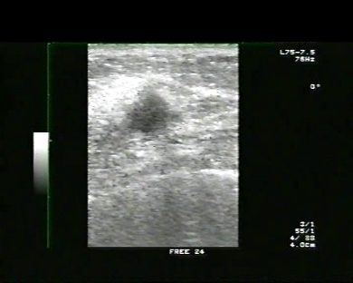
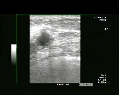
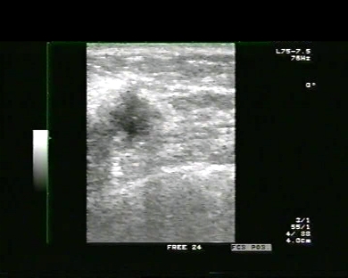
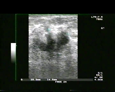
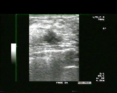
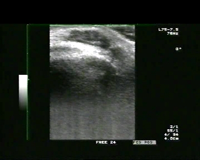
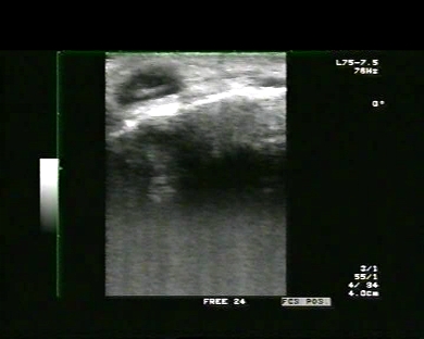
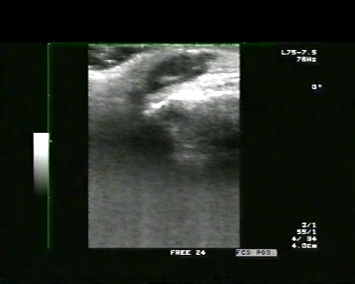
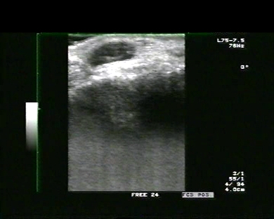
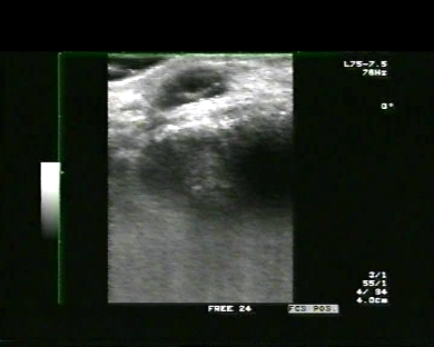
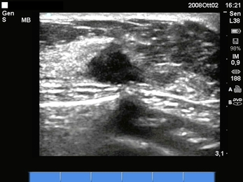
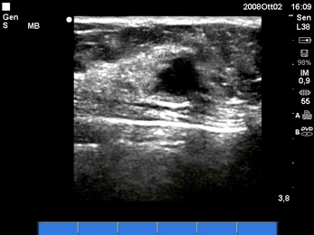
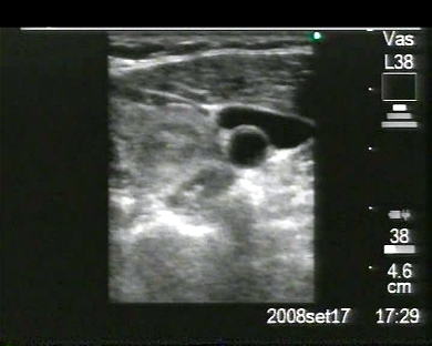
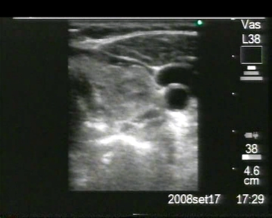
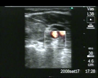
Inclusion date: 12 /12/2008Ultrasound made on: 03/12/2008 Instrument: Esaote Megas Patient's age: F 76 years old The ultrasound is carried out following a mammography which highlights a nodule in the left breast. The images and the video show an oval and hypoechoic formation, with irregular, angular margins and small calcifications within the nodule. It does not, however, show a vascularization. The dimensions of the formation is 17mm x 15mm, and it is located in the external upper quadrant of the left breast. The above mentioned ultrasound characteristics lead towards the diagnosis of breast cancer. In collaboration with: Dr. Pietro Vitali - Jesi (AN) Presentation: Dr. Massimo Dolciotti - Ancona Digital processing: Andrea Dini - Ancona
|
|
|
|
|
|
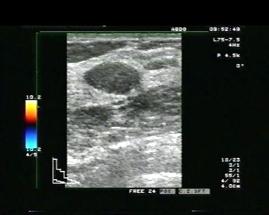
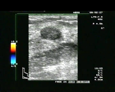
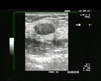
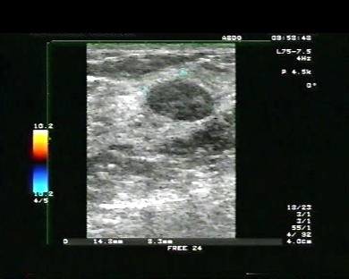
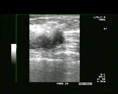
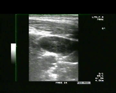
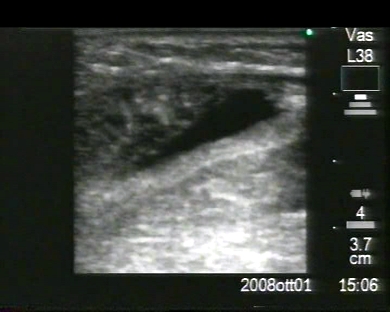
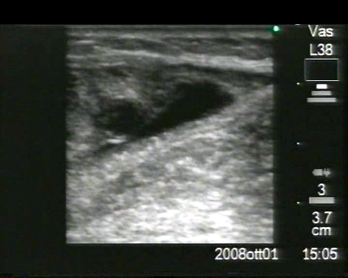
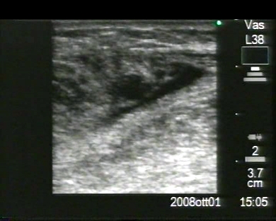
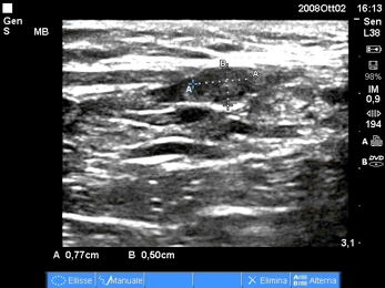
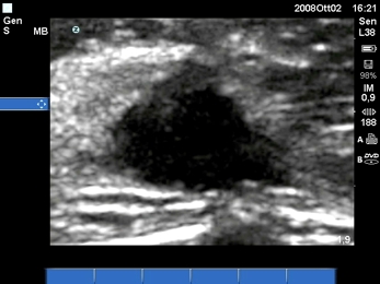
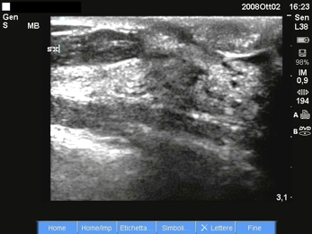
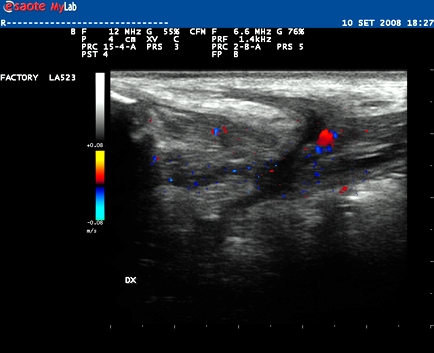
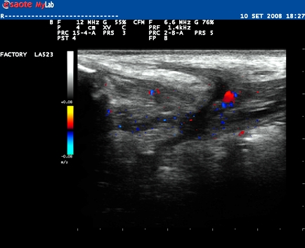
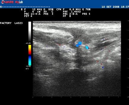
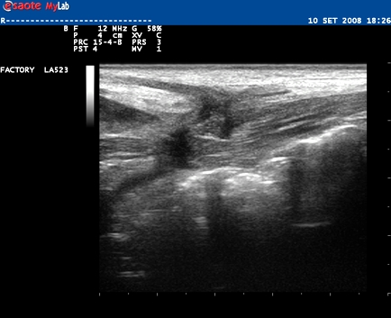
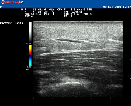
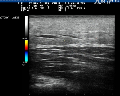
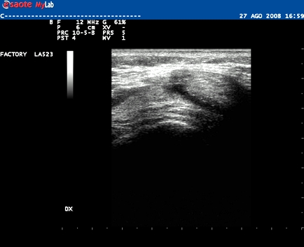
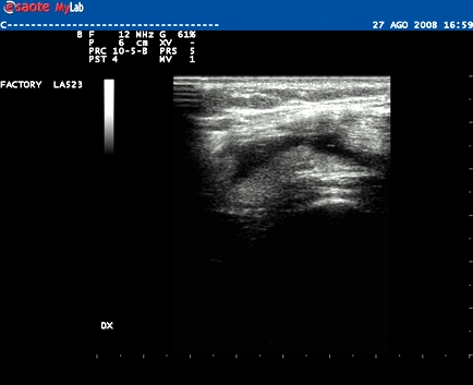
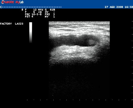
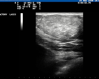
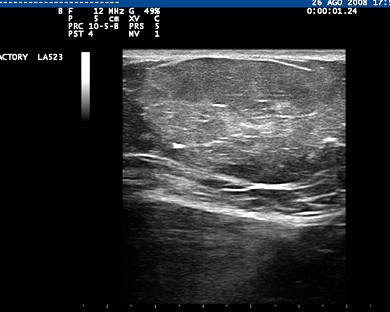
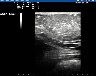
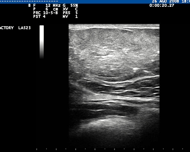
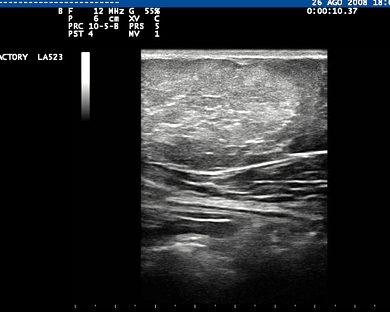
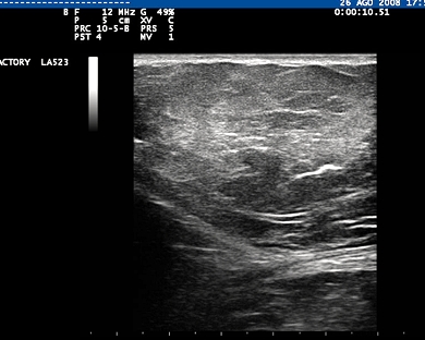
Inclusion date: 10 /12/2008Ultrasound made on: 10/11/2008 Instrument: Toshiba 380A Patient's age: F 43 years old The ultrasound examination is carried out as a follow up for a breast nodule located in the external upper quadrant of the right breast. The images and the video show a nodule with regular and clean margins, acoustic shadows on the side and a reinforced back wall. The nodule is well-defined by the surrounding glandular tissue, it is painless, mobile and little vascularised, and it is 16 x 8 mm in size. The above ultrasound characteristics lead towards the diagnosis of a fibroadenoma. Presentation: Dr. Massimo Dolciotti - Ancona Digital processing: Andrea Dini - Ancona
|
|
|
|
|
|
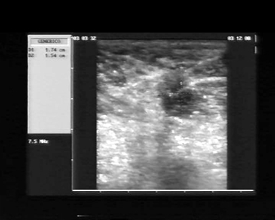
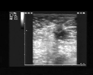
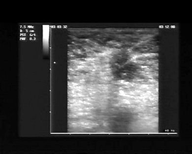
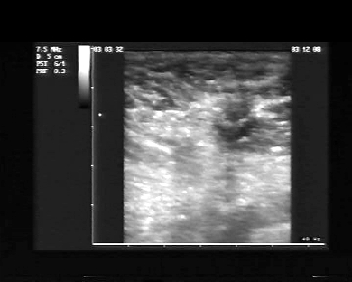
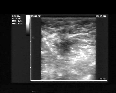
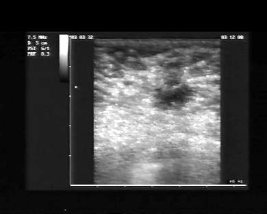
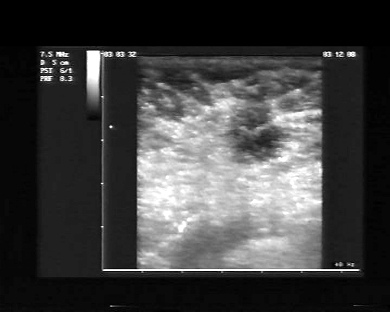
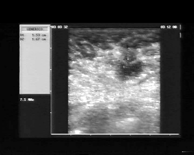
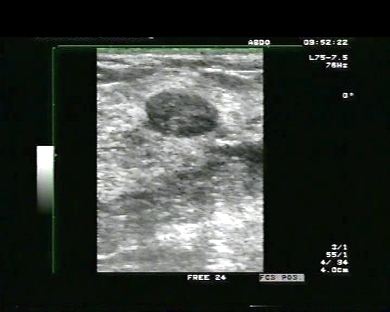
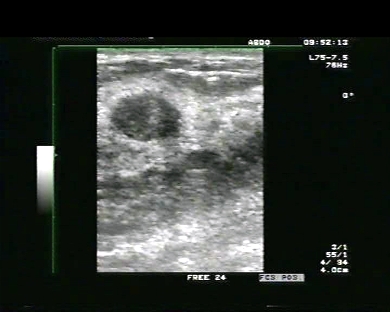
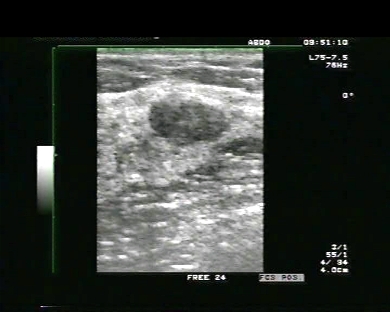
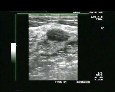
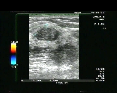
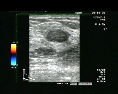
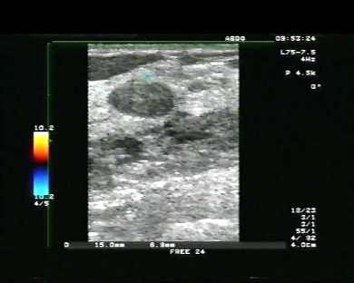
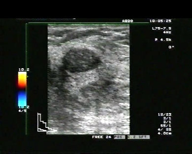
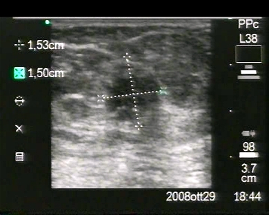
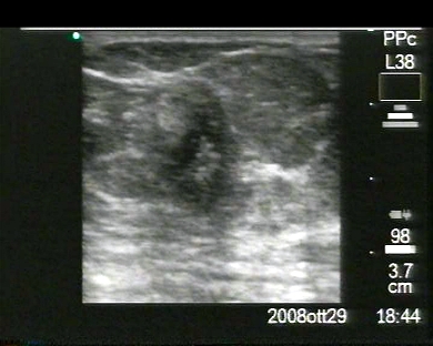
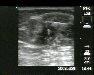
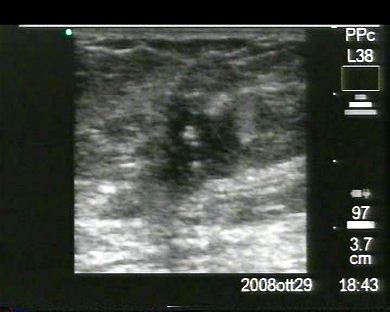
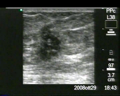
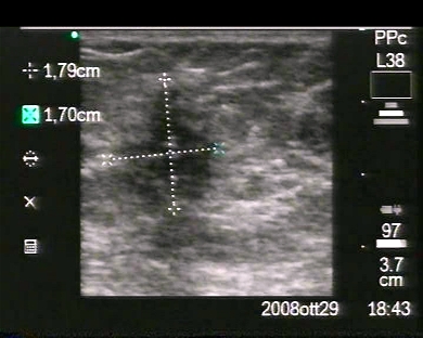
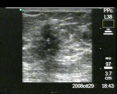
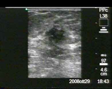
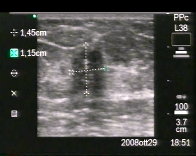
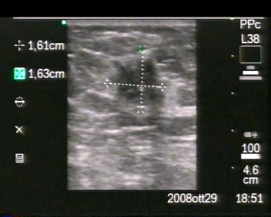
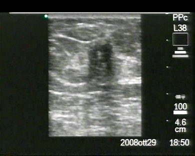
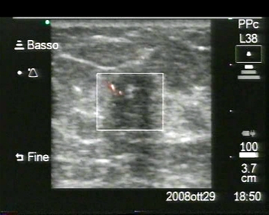
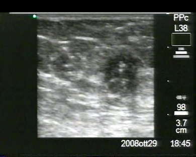
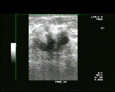
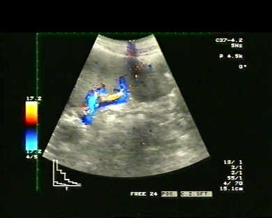
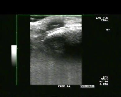
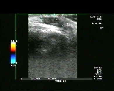
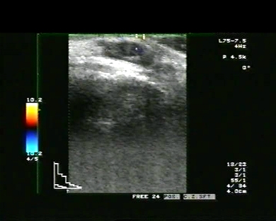
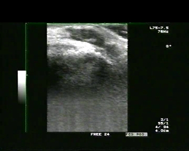
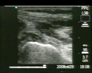
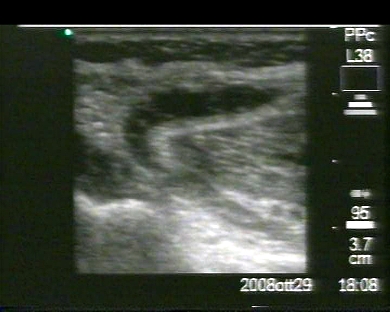
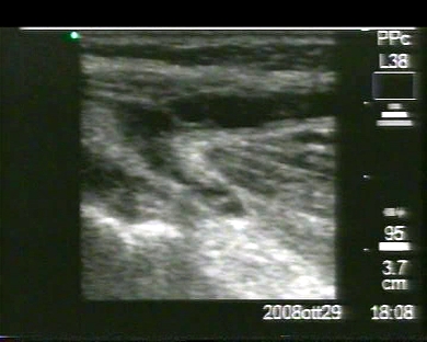
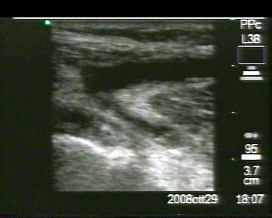
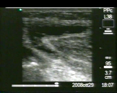
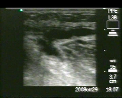
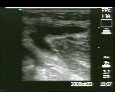
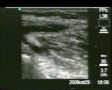
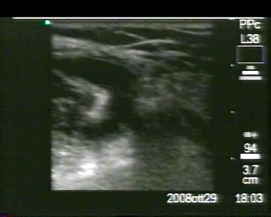
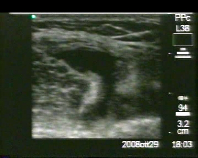
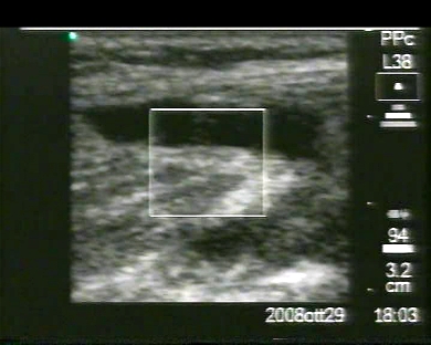
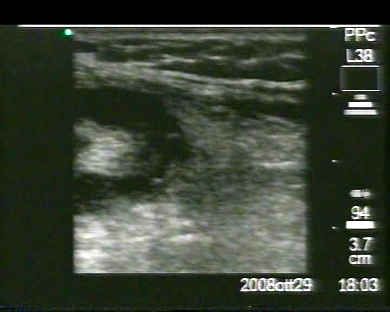
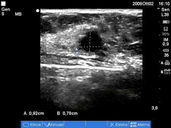
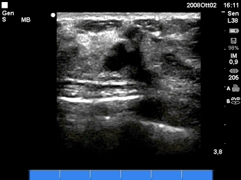
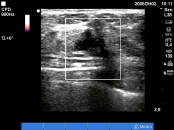
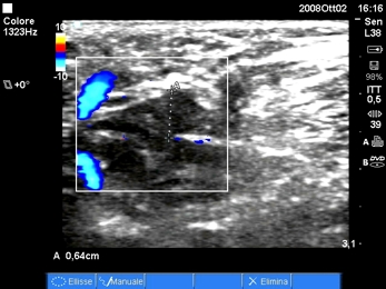
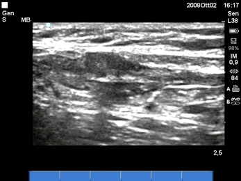
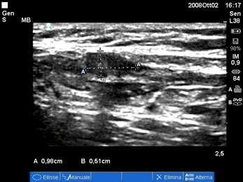
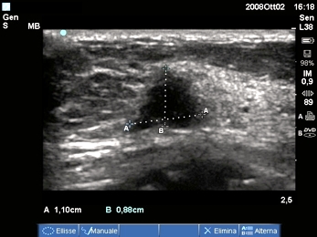
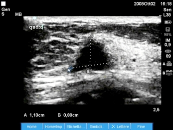
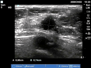
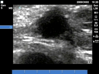
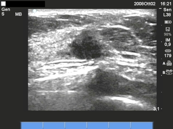
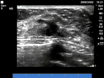
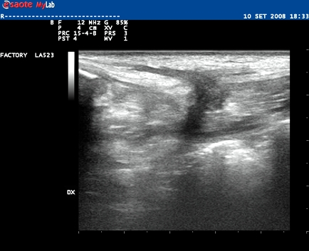
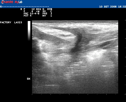
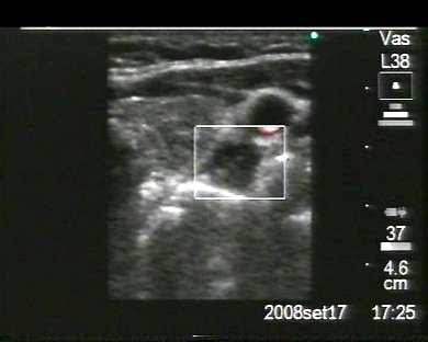
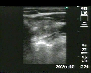
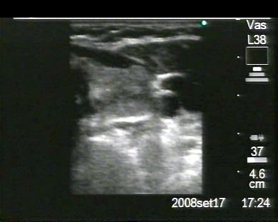
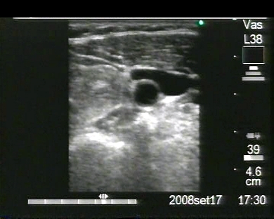
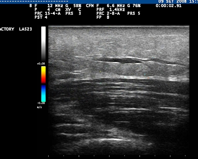
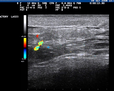
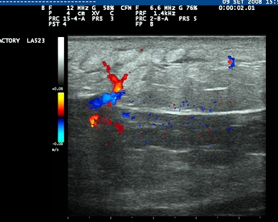
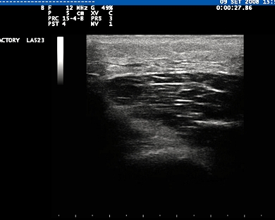
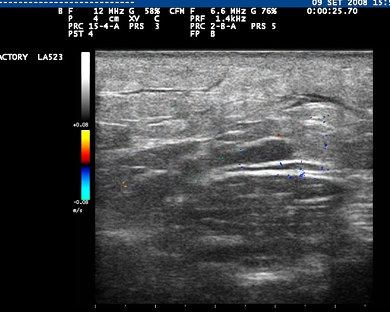
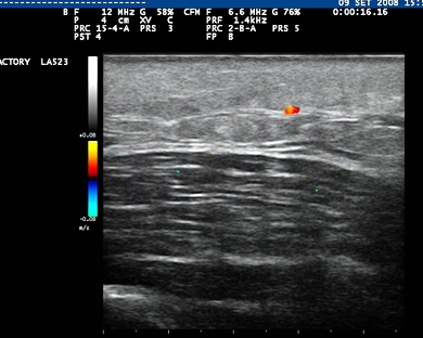
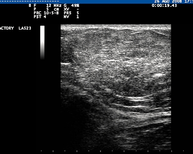
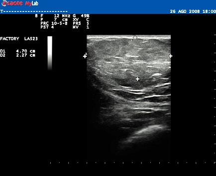
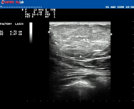
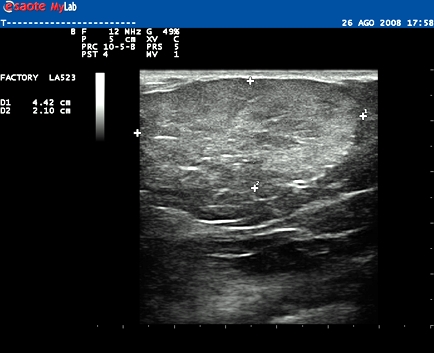
Inclusion date: 05 /12/2008Ultrasound made on: 29/10/2008 Instrument: Sonosite Patient's age: F 76 years old The ultrasound examination is carried out as a follow-up of a breast neoplasia, which has already undergone a right quadrantectomy. The images and the video show a nodule in the upper quadrant of the left breast. The nodule, of 17 x 16mm in size, has irregular margin and minimum vascular signals. In collaboration with: Dr. Pietro Vitali - Jesi (AN) Presentation: Dr. Massimo Dolciotti - Ancona Digital processing: Andrea Dini - Ancona
|
|
|
|
|
|
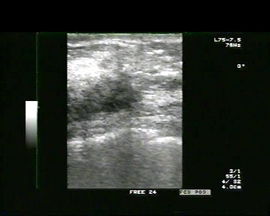
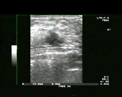
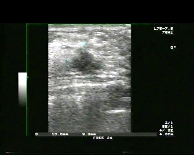
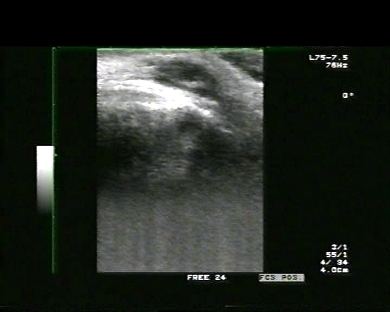
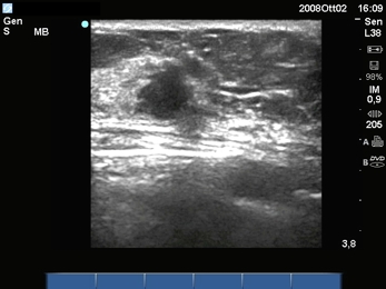
Inclusion date: 01 /12/2008Ultrasound made on: 14/10/2008 Instrument: Toshiba 380A Patient's age: F 71 years old The ultrasound examination is carried out following a hard swelling of the right breast. The images and video show a hypoechoic nodule with irregular margins and no vascular signs. It measures 13 x 9.8 mm. The hystologic examination of the mastectomy shows infiltrating breast cancer with ductal and lobular aspects (mixed type) not very differentiated (grade 3 according to Elston-Ellis) and associated to a minimal intraductal component of an intermediate grade. Receptors for the Estrogens: Positive 95%. Receptors for the Progesterone: Positive 35%. Proliferative activity (Mib1): High 35%. Expression of p53: Negative 3%. Presentation: Dr. Massimo Dolciotti - Ancona Digital processing: Andrea Dini - Ancona
|
|
|
|
|
|
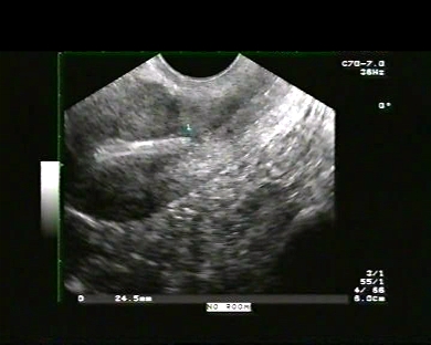
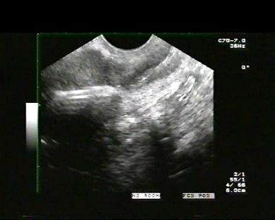
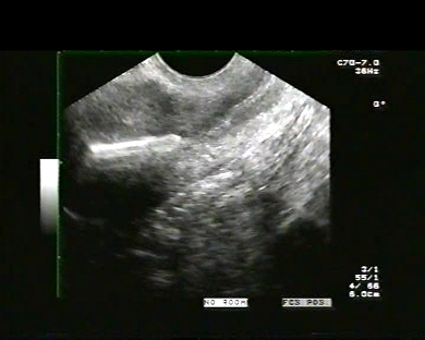
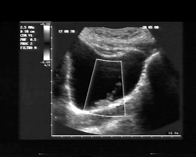
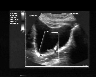
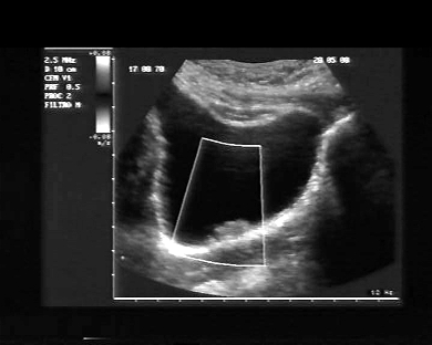
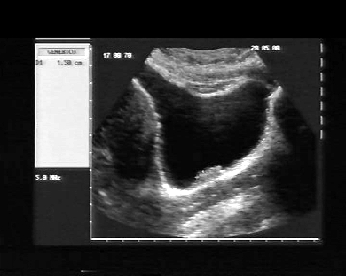
Inclusion date: 26 /11/2008Ultrasound made on: 30/09/2008 Instrument: Toshiba 380A Patient's age: F 28 years old The ultrasound examination is carried out following pain in the lower abdomen. The images and the video show the anteverted uterus with a regular echo-structure and morphovolumetry, and a regularly positioned intra-uterine device (IUD). The right and left ovaries have a regular echo-morphovolumetry. Presentation: Dr. Massimo Dolciotti - Ancona Digital processing: Andrea Dini - Ancona
|
|
|
|
|
|
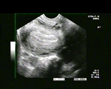
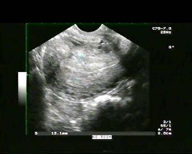
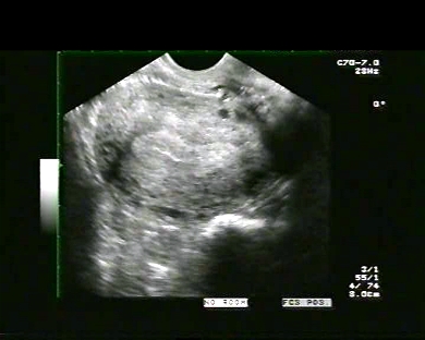
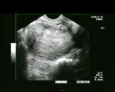
Inclusion date: 26 /11/2008Ultrasound made on: 18/11/2008 Instrument: Toshiba 380A Patient's age: F 18 years old The ultrasound examination is carried out to study the pelvis from a suprapubic angle. The images and the video show the retroverted uterus with a regular echo-structure and morphovolumetry. The uterus is in the menstrual phase and there is evidence of an internal tampon in the vaginal canal. The endometrium is scarcely visualised. Presentation: Dr. Massimo Dolciotti - Ancona Digital processing: Andrea Dini - Ancona
|
|
|
|
|
|
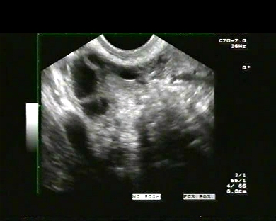
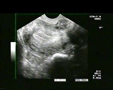
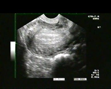
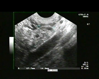
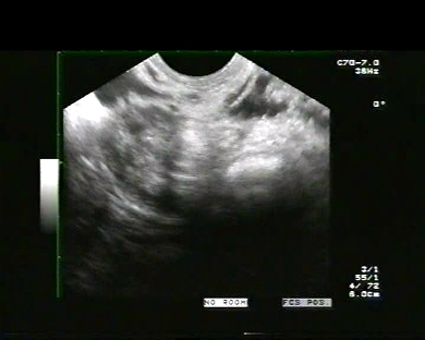
Inclusion date: 24 /11/2008Ultrasound made on: 01/10/2008 Instrument: Toshiba 380A Patient's age: F 24 years old The ultrasound examination is carried out for a detailed study of the uterus, of the endometrium and of the ovaries. The images and the video show the anteverted uterus with a regular echo-structure and morphovolumetry. The endometrium is in a phase of secretion and is 13-14mm thick. The ovaries have regular dimensions and echostructure. Presentation: Dr. Massimo Dolciotti - Ancona Digital processing: Andrea Dini - Ancona
|
|
|
|
|
|
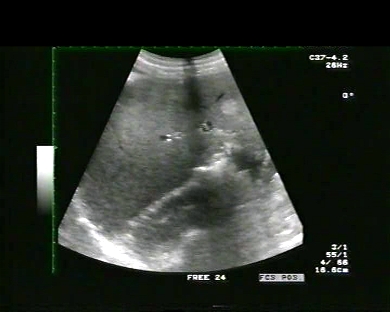
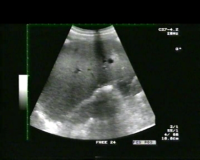
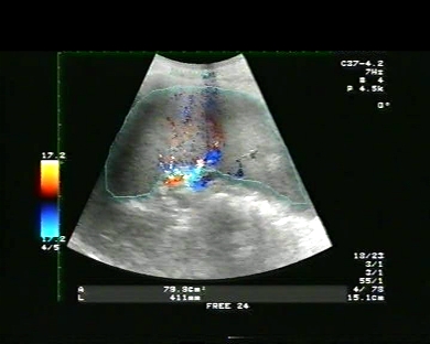
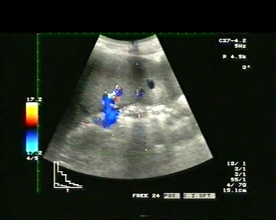
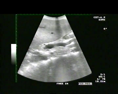
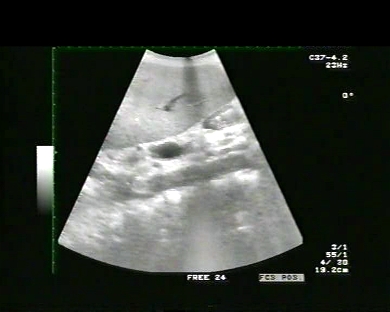
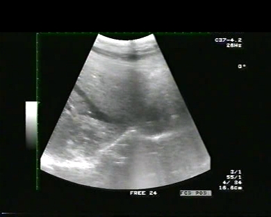
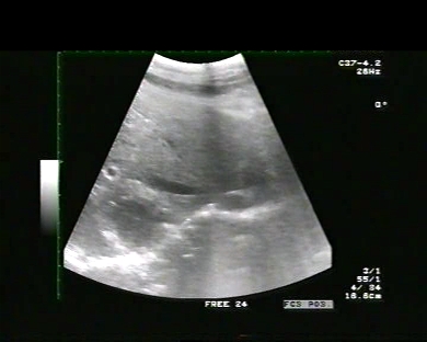
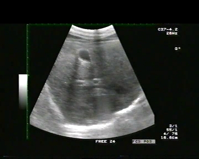
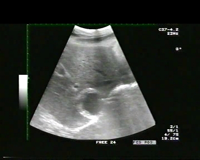
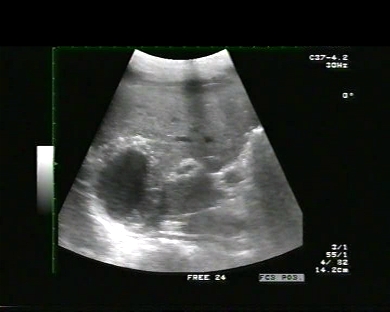
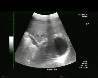
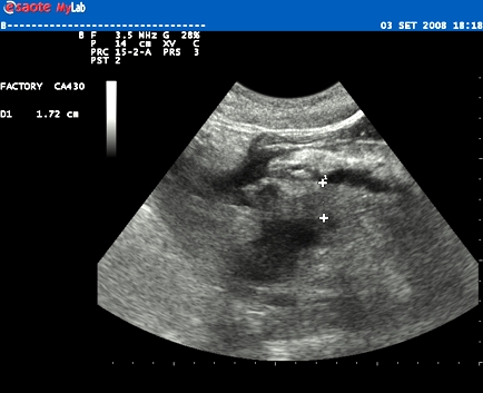
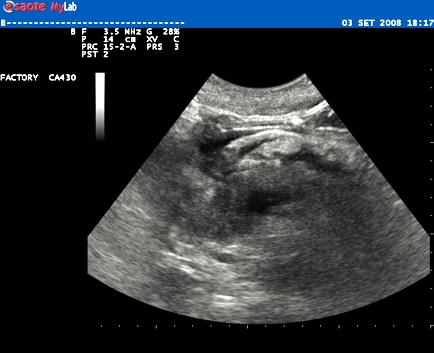
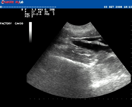
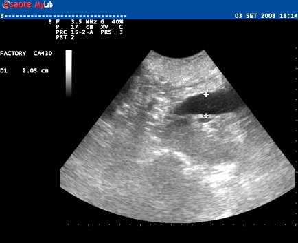
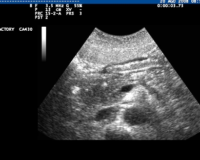
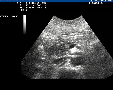
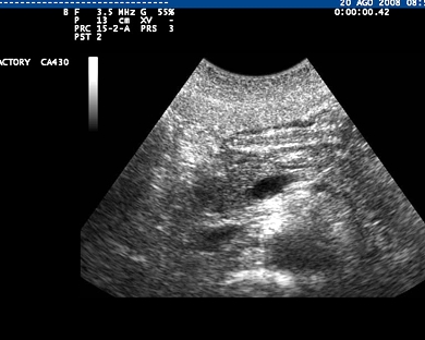
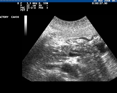
Inclusion date: 21 /11/2008Ultrasound made on: 20/10/2008 Instrument: Toshiba 380A Patient's age: F 50 years old The ultrasound examination is carried out as a follow-up of a chronic hepatopathy. The images and the video show the spleen with an increased volume and a section area of 79 square cm. In the inferior pole there is evidence of a small cyst and an angioma measuring 15x11mm. Presentation: Dr. Massimo Dolciotti - Ancona Digital processing: Andrea Dini - Ancona
|
|
|
|
|
|
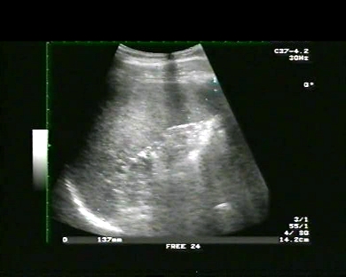
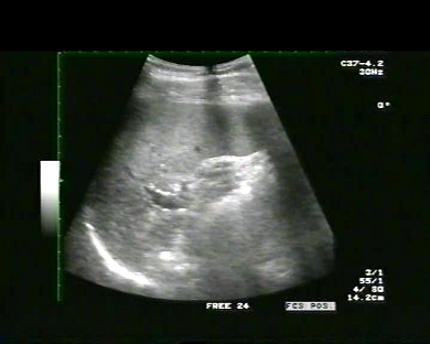
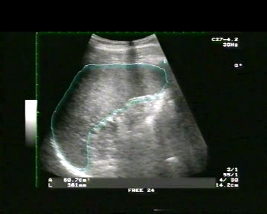
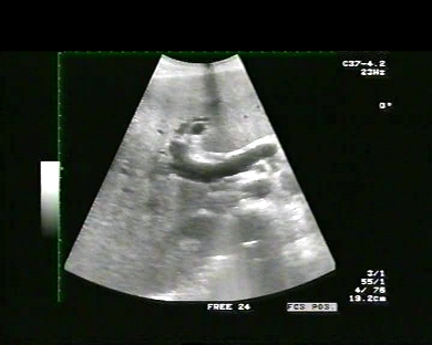
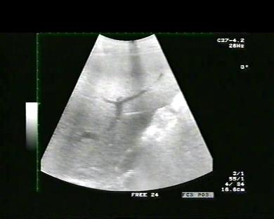
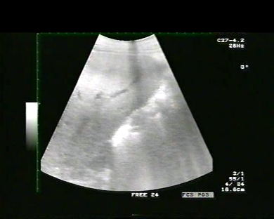
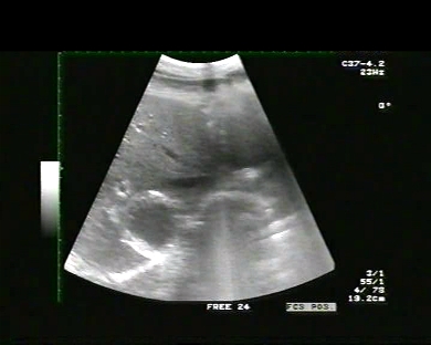
Inclusion date: 19 /11/2008Ultrasound made on: 23/04/2008 Instrument: Toshiba 380A Patient's age: M 18 years old The ultrasound examination is carried out following slight temperature, bigger lymph nodes in the neck and exanthema. The images and the video show lymph nodes which are increased in volume and are located at the back of the nucha, on the fifth level of the surgical division of the lymph nodes of the neck. They also show a splenomegaly with a section area of 60 square cm and bipolar diameter of 137 mm. The clinic and the scan lead towards the diagnosis of rubella. Presentation: Dr. Massimo Dolciotti - Ancona Digital processing: Andrea Dini - Ancona
|
|
|
|
|
|
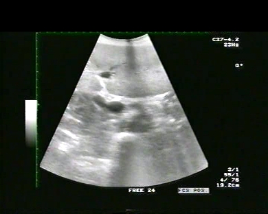
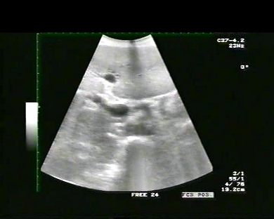
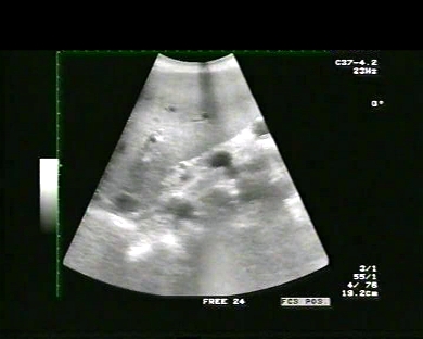
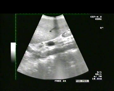
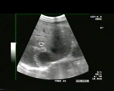
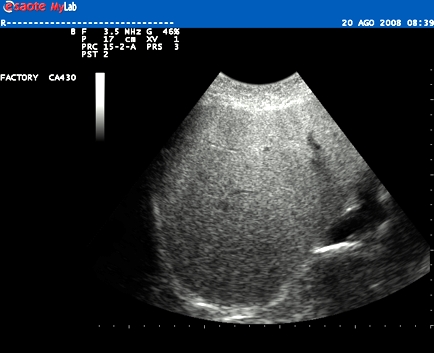
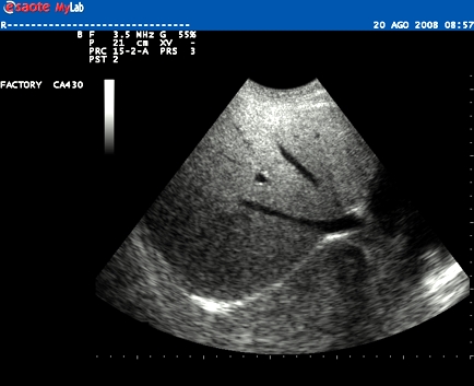
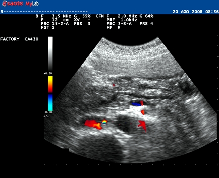
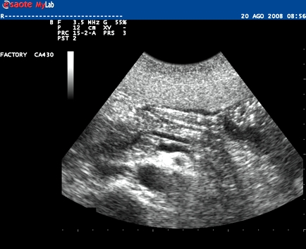
Inclusion date: 14 /11/2008Ultrasound made on: 20/10/2008 Instrument: Toshiba 380A Patient's age: F 50 years old The ultrasound examination is carried out following a chronic, alcoholic hepatopathy in a patient diagnosed as HIV positive. The images and the video show the liver with a morphovolumetry above normal, irregular margins and obtuse angles, medium-sized and with widespread steatosis and a portal vein with increased calibre (15 mm –V.N. < 12 mm). Presentation: Dr. Massimo Dolciotti - Ancona Digital processing: Andrea Dini - Ancona English translation: Prof.ssa Federica Sturani - England
|
|
|
|
|
|
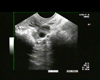
Inclusion date: 14 /11/2008Ultrasound made on: 11/01/2008 Instrument: Toshiba 380A Patient's age: F 17 years old The ultrasound examination is carried out following pain in the lower abdomen. The images and the video show the anteverted uterus with a regular echo-structure and morphovolumetry, and the endometrium in a proliferative phase and with an 11 mm-thick trilaminar aspect. Well evident is the vaginal canal which is extended due to the presence of abundant faecal material in the rectal ampulla. The faecal material is identified by the hyperechoic aspect. The images also show a mature ovarian follicle in the right ovary. The bladder is modestly full and with regular walls. Presentation: Dr. Massimo Dolciotti - Ancona Digital processing: Andrea Dini - Ancona English translation: Prof.ssa Federica Sturani - England
|
|
|
|
|
|
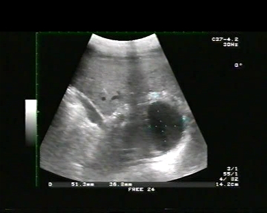
Inclusion date: 12 /11/2008Ultrasound made on: 04/11/2008 Instrument: Toshiba 380A Patient's age: M 26 years old The ultrasound examination is carried out as a follow-up after 30 days from the abdominal trauma with cyst lesion, which is located in the eighth hepatic segment and measures 47 x 35 mm. The images and the video show the anechoic image with irregular margins and with a back acoustic enhancement. Presentation: Dr. Massimo Dolciotti - Ancona Digital processing: Andrea Dini - Ancona English translation: Prof.ssa Federica Sturani - England
|
|
|
|
|
|
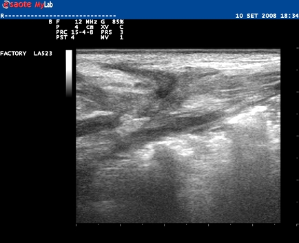
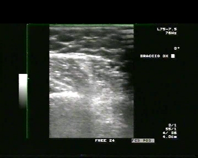
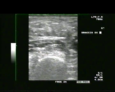
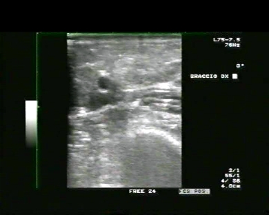
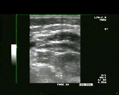
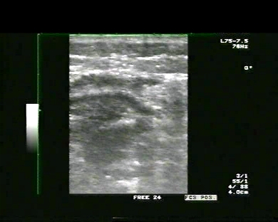
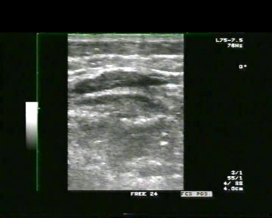
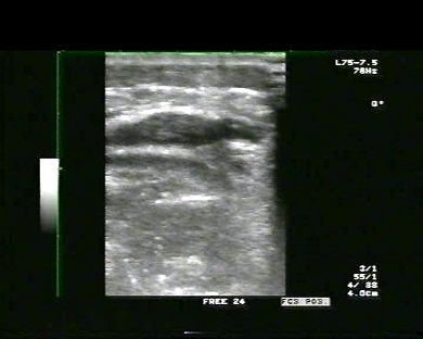
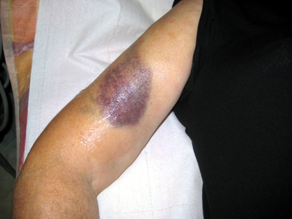
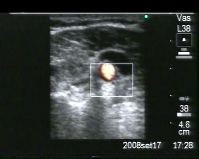
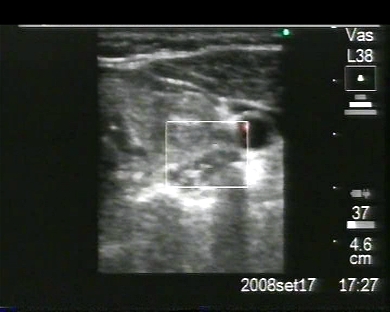
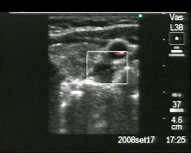
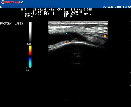
Inclusion date: 10 /11/2008Ultrasound made on: 01/10/2008 Instrument: SonositePatient's age: M 53 years old The ultrasound examination is carried out following swelling and pain in the distal region of the right leg. The images and the video show in the distal site the myotendinous lesion to the left vastus medialis. In collaboration with: Dr. Daniele Lenti - Jesi (AN) Presentation: Dr. Massimo Dolciotti - Ancona Digital processing: Andrea Dini - Ancona English translation: Prof.ssa Federica Sturani - England
|
|
|
|
|
|
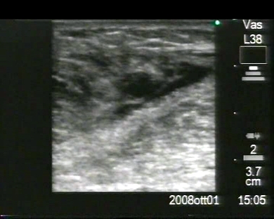
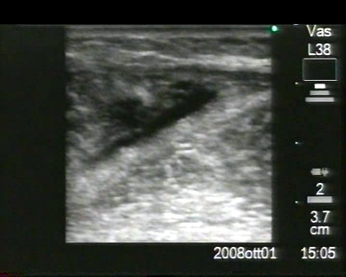
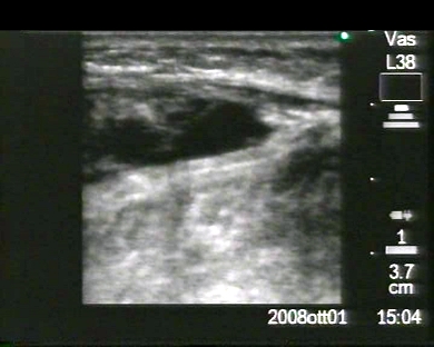
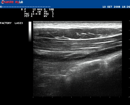
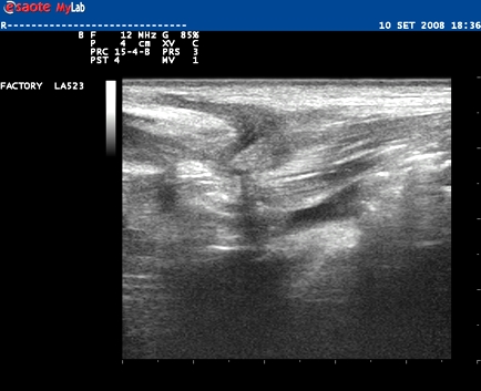
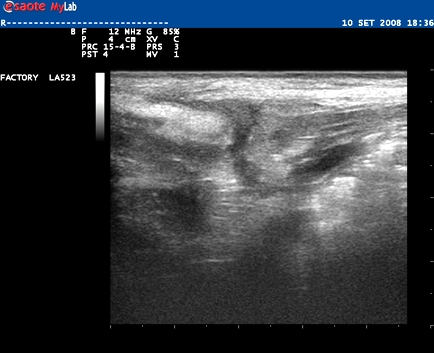
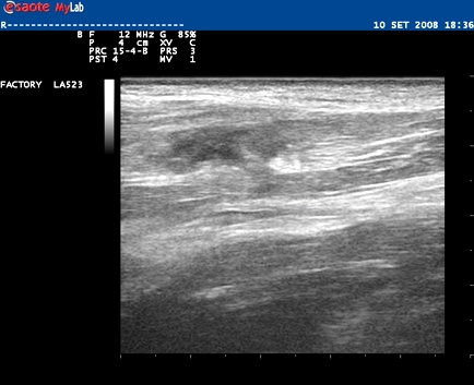
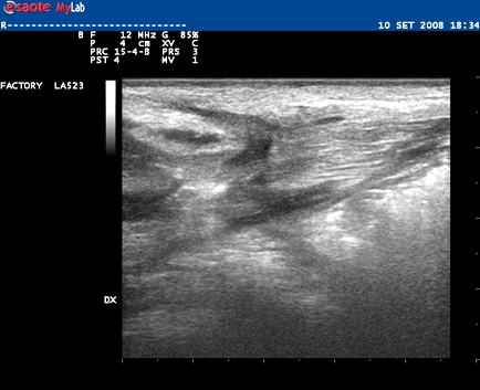
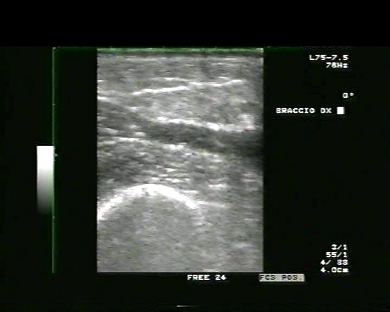
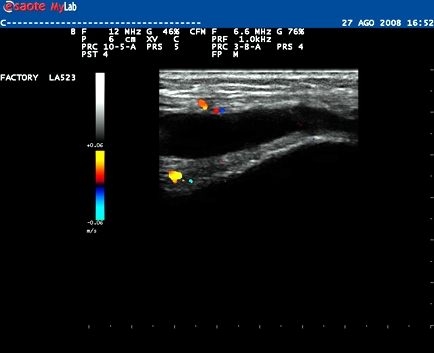
Inclusion date: 10 /11/2008Ultrasound made on: 29/10/2008 Instrument: SonositePatient's age: M 60 years old The ultrasound examination is carried out following swelling and pain in the medial region of the right leg. The images and the video show the myotendinous lesion of the right vastus medialis. In collaboration with: Dr. Daniele Lenti - Jesi (AN) Presentation: Dr. Massimo Dolciotti - Ancona Digital processing: Andrea Dini - Ancona English translation: Prof.ssa Federica Sturani - England
|
|
|
|
|
|
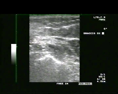
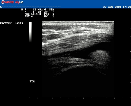
Inclusion date: 03 /11/2008Ultrasound made on: 01/10/2008 Instrument: SonositePatient's age: F 58 years old The ultrasound examination is carried out following painful swelling in the right popliteal fossa. The images and the video show the liquid cyst, or Baker’s cyst, which is the collection of fluid in the medial and semi-membranal gastrocnemius muscle. Presentation: Dr. Massimo Dolciotti - Ancona Digital processing: Andrea Dini - Ancona English translation: Prof.ssa Federica Sturani - England
|
|
|
|
|
|
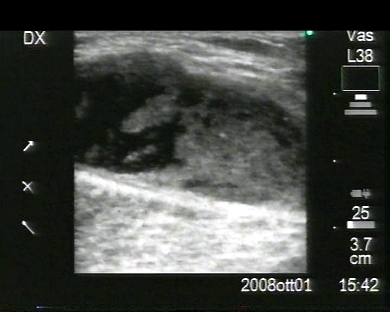
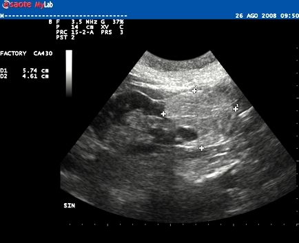
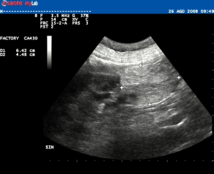
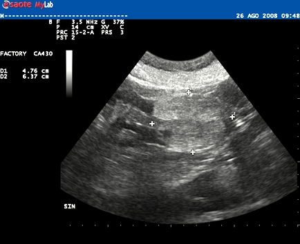
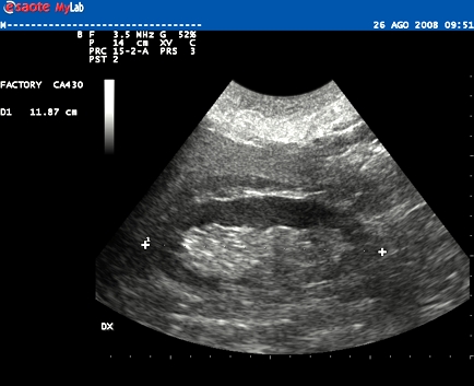
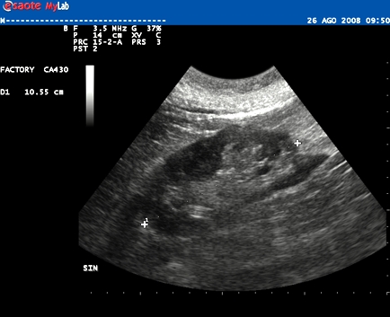
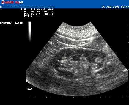
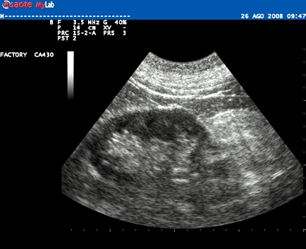
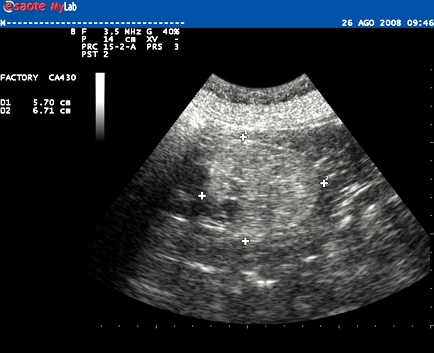
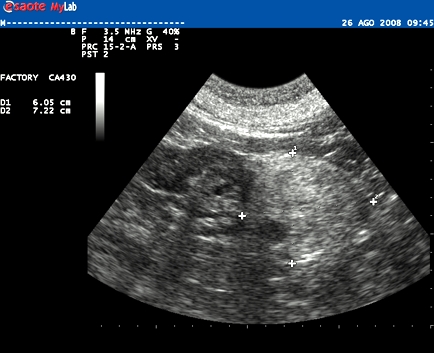
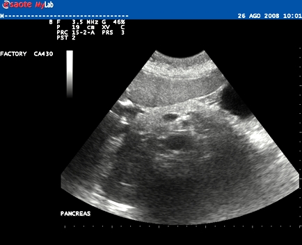
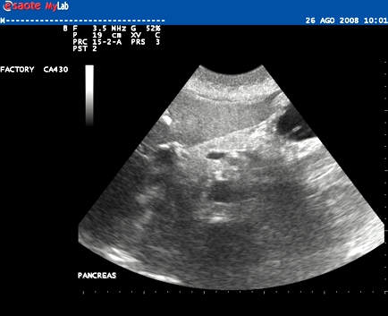
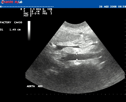
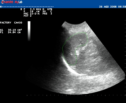
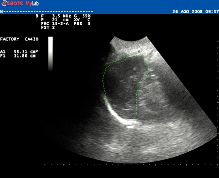
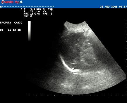
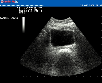
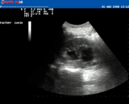
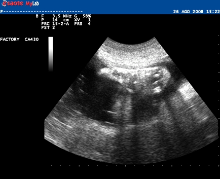
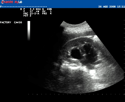
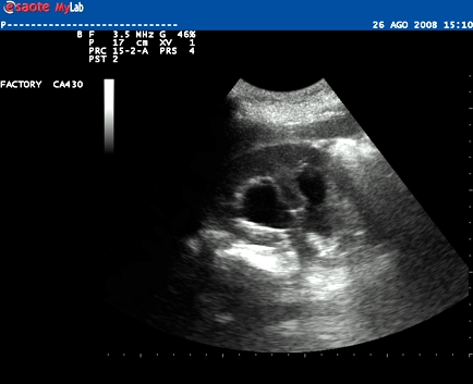
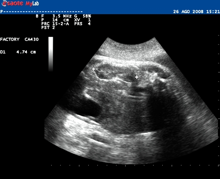
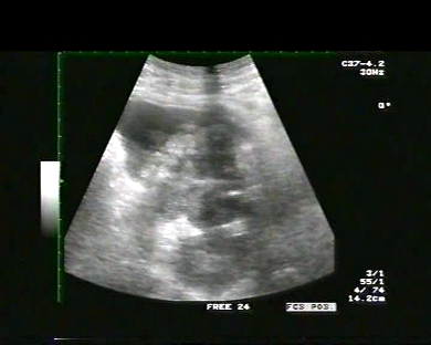
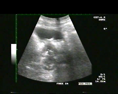
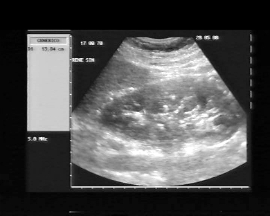
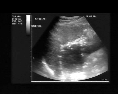
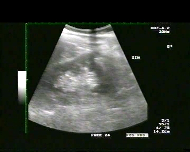
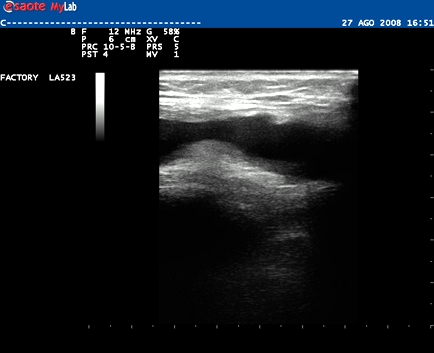
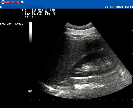
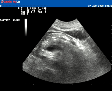
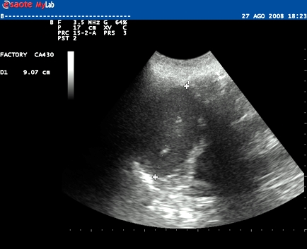
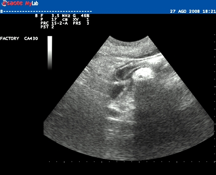
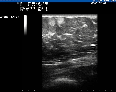
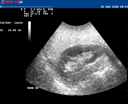
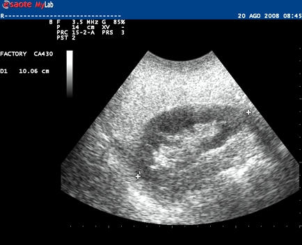
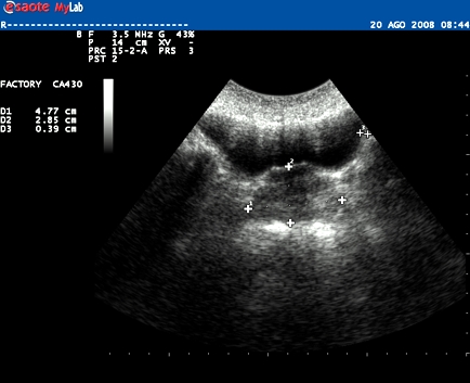
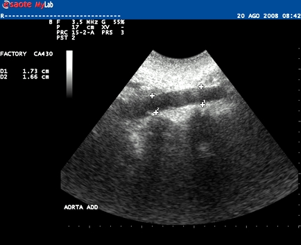
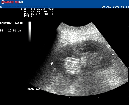
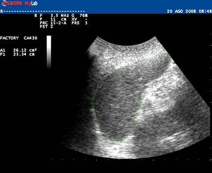
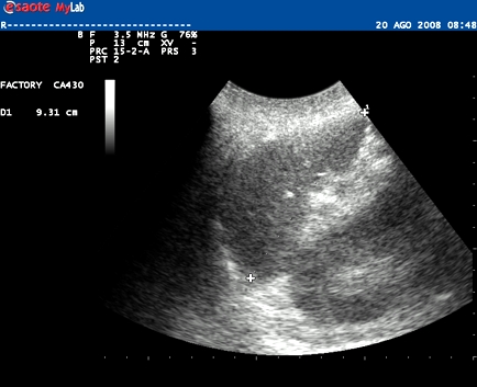
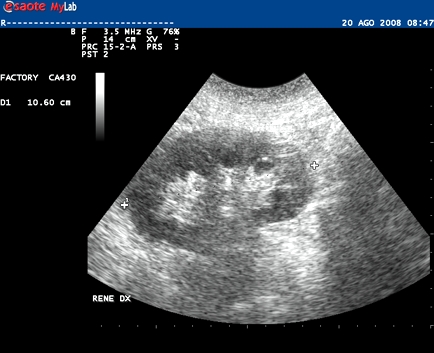
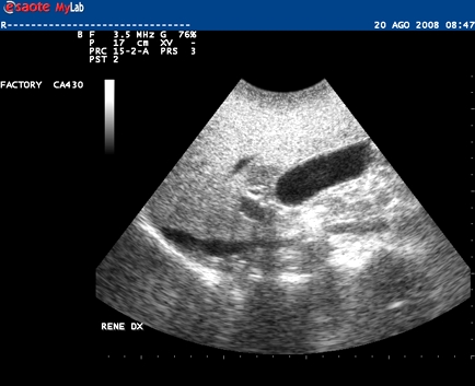
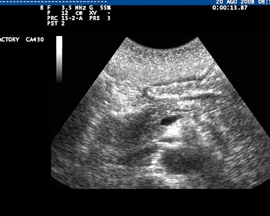
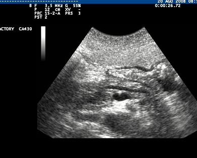
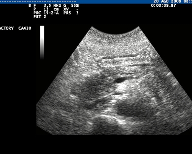
Inclusion date: 31 /10/2008Ultrasound made on: 26/08/2008 Instrument: Esaote MyLab 25Patient's age: F 75 years old The ultrasound examination is carried out as a follow-up of a lipoma which is located in the inferior pole of the left kidney and which is found to be of unchanged dimension from the check-up of the previous year. Presentation: Dr. Massimo Dolciotti - Ancona Digital processing: Andrea Dini - Ancona English translation: Prof.ssa Federica Sturani - England
|
|
|
|
|
|
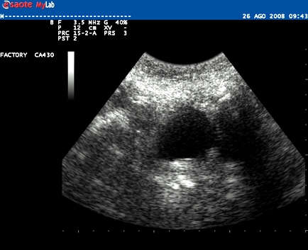
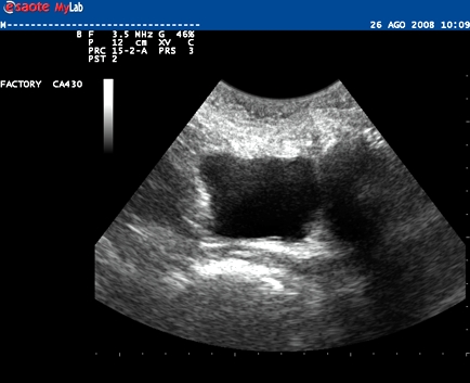
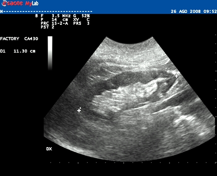
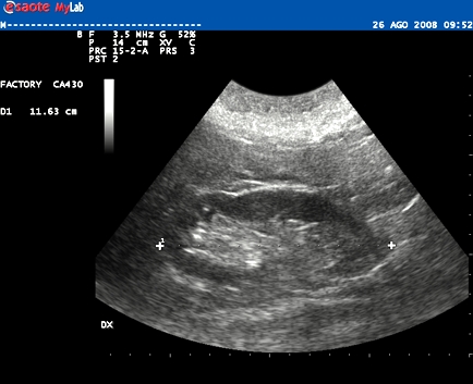
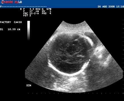
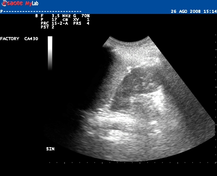
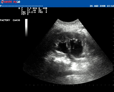
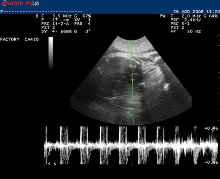
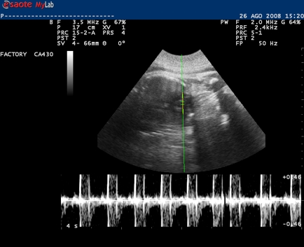
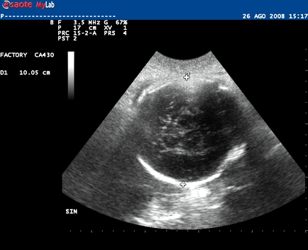
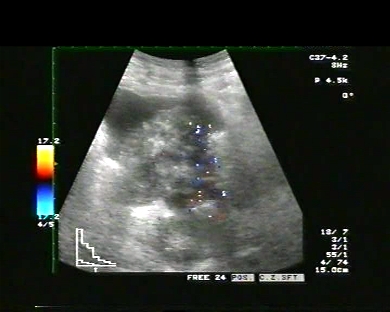
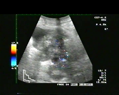
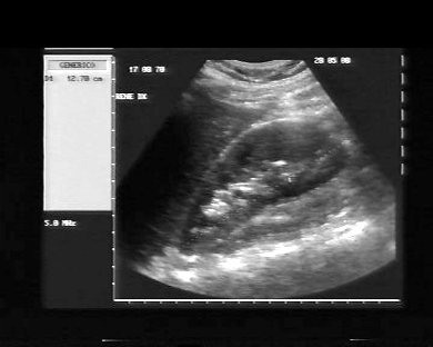
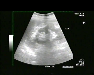
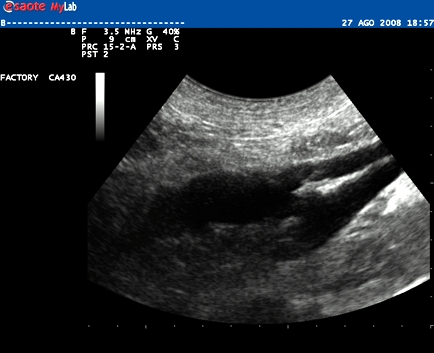
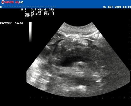
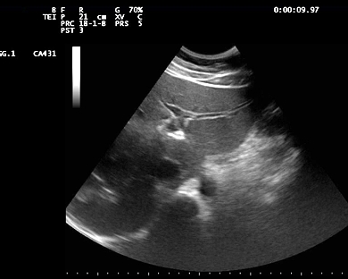
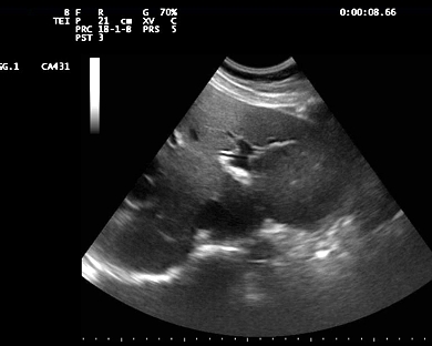
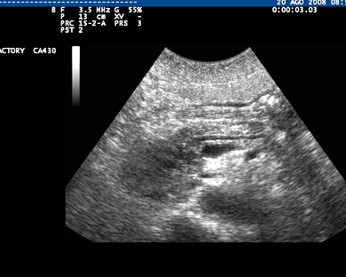
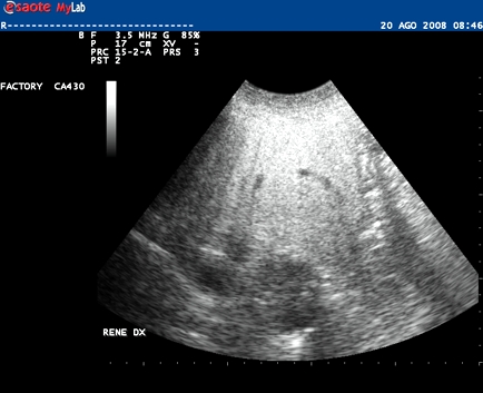
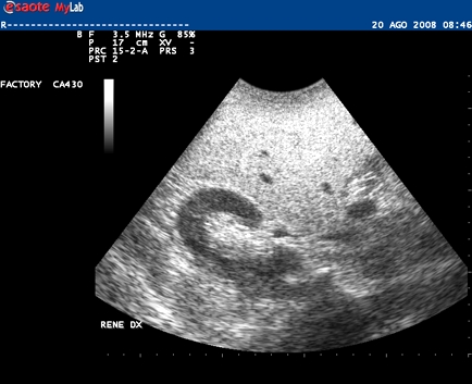
Inclusion date: 29 /10/2008Ultrasound made on: 26/08/2008 Instrument: Esaote MyLab 25Patient's age: F 22 years old The ultrasound examination is carried out as a follow-up of a hydronephrosis in the right kidney in the final stage of a pregnancy. The images and the video show that the medium-sized hydronephrosis in the right kidney has slightly increased from the previous check-up. Presentation: Dr. Massimo Dolciotti - Ancona Digital processing: Andrea Dini - Ancona English translation: Prof.ssa Federica Sturani - England
|
|
|
|
|
|
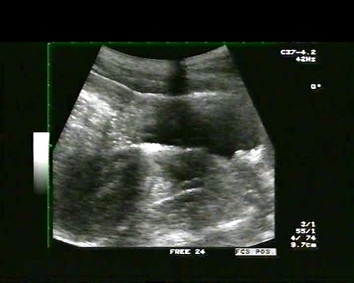
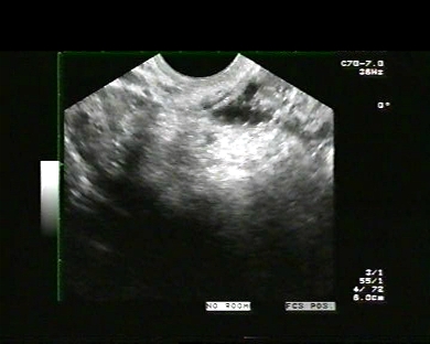
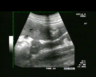
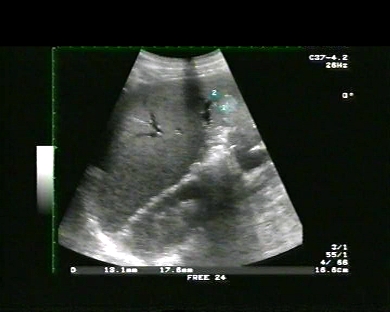
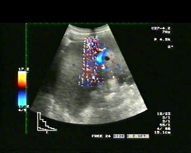
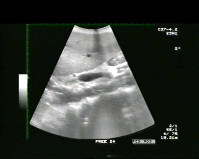
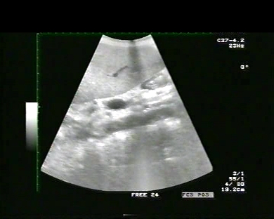
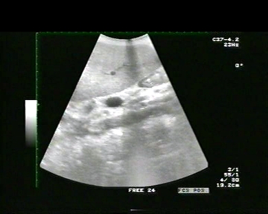
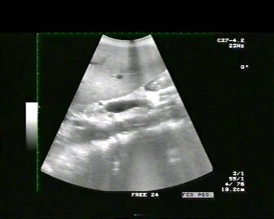
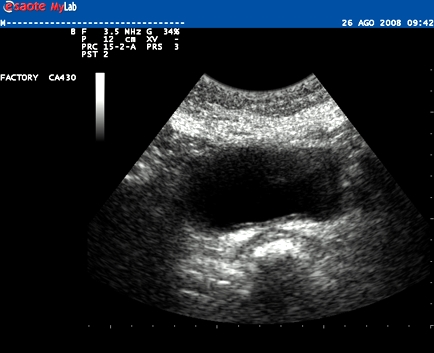
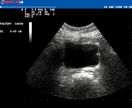
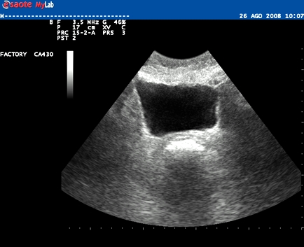
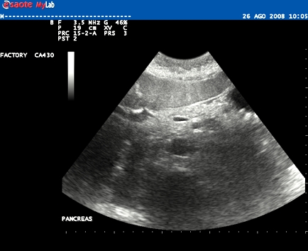
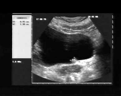
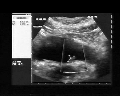
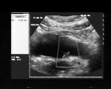
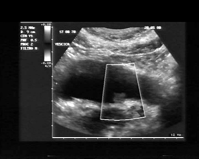
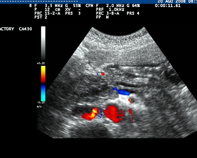
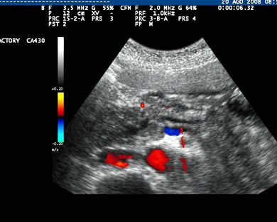
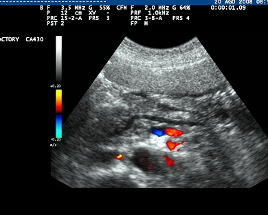
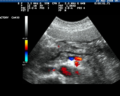
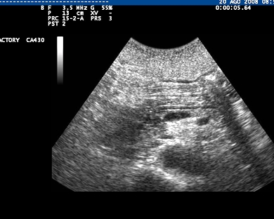
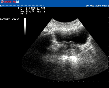
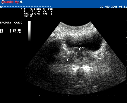
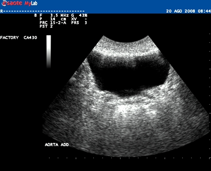
Inclusion date: 22/ 10/2008Ultrasounds made on: 22/09/2008 Instrument: Toshiba 380APatient's age: M 51 years old The patient undergoes an ultrasound examination following pains at the lower abdomen 22 months after a surgical operation of left hemicolectomy required by an infiltrating adenocarcinoma. The images and the video show the recurrence in the rectum-vesical space, with an initial infiltration in the vesical wall. Presentation: Dr. Massimo Dolciotti - Ancona - Italy Digital processing: Andrea Dini - Ancona - Italy English translation: Prof.ssa Federica Sturani - England
|
|
|
|
|
|
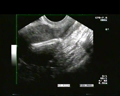
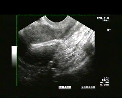
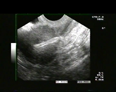
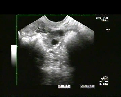
Inclusion date: 20/ 10/2008Ultrasounds made on: 29/09/2008 Instrument: Toshiba 380APatient's age: F 32 years old The patient undergoes an ultrasound examination in the upper-pubis using a 3.5 Mhz convex probe, 48 hours after a surgical LEEP conisation. The conisation was required by a carcinoma in the uterine cervix caused by papillomavirus. The images and the video show the anteverted uterus, with a thickening of the isthmus region of the uterus and an alteration of its normal profile. Presentation: Dr. Massimo Dolciotti - Ancona - Italy Digital Processing: Andrea Dini - Ancona - Italy English translation: Prof.ssa Federica Sturani - England
|
|
|
|
|
|
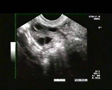
Inclusion date: 17/ 10/2008Ultrasounds made on: 15/12/2006 - 02/01/2007 Instrument: Toshiba 380A Patient's age: F 29 years old Within the context of women infertility, the upper-pubis ultrasound examination can help a woman with regular menstruation cycles to become pregnant by analysing the maturation of the ovarian follicle and, at the same time, visualising the endometrium, which in a periovular phase becomes trilaminar. This ultrasound case study shows a predominant follicle of approx. 17-18 mm in the right ovary and a trilaminar endometrium, both of which are a sign of fertile days for the woman. Indeed, the following ultrasound check-up, which was carried out 18 days after, shows the patient’s initial stage of pregnancy. (Last menstruation: 20/11/2006; betaHCG on 02/01/2007: 20,735 ui/l). Presentation: Dr. Massimo Dolciotti - Ancona - Italy Digital processing:: Andrea Dini - Ancona - Italy English translation: Prof.ssa Federica Sturani - England
|
|
|
|
|
|
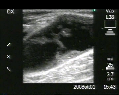
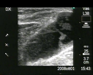
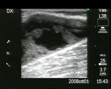
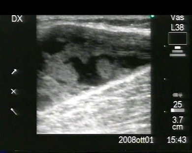
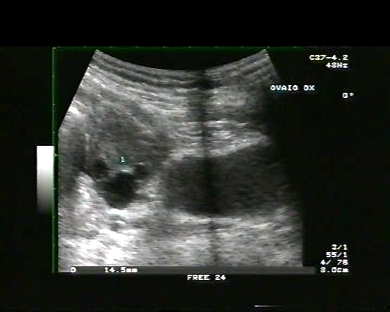
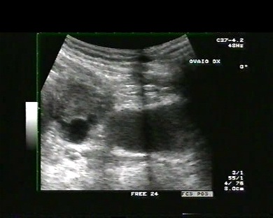
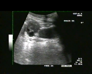
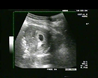
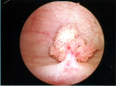
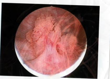
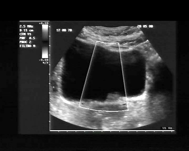
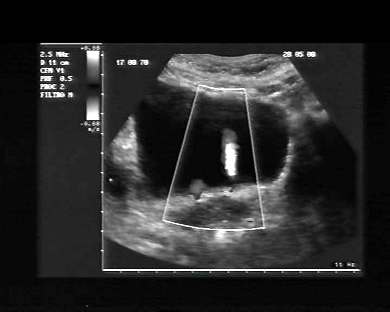
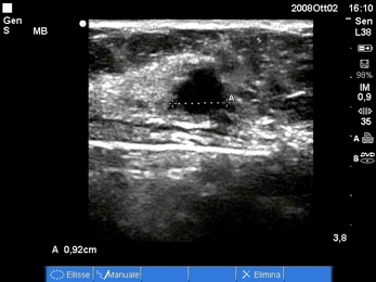
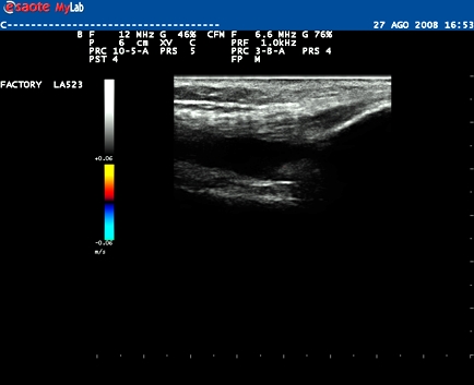
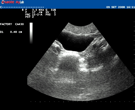
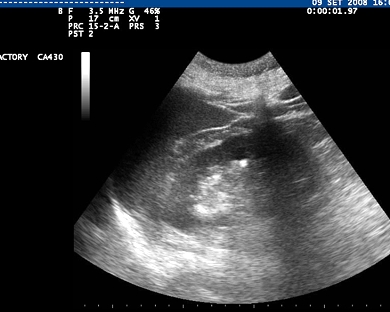
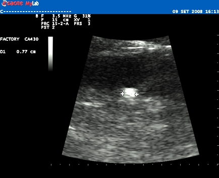
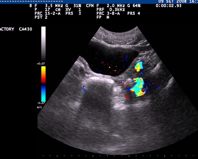
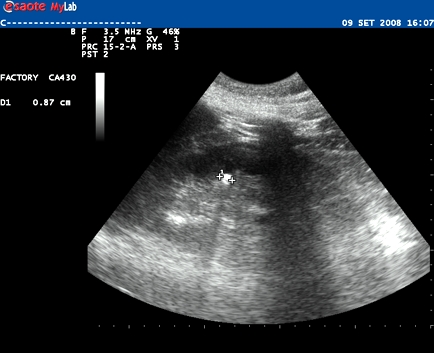
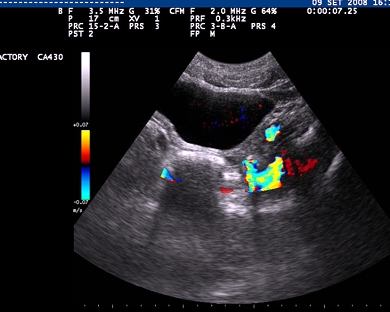
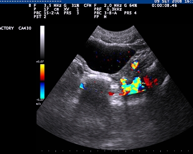
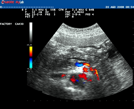
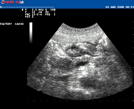
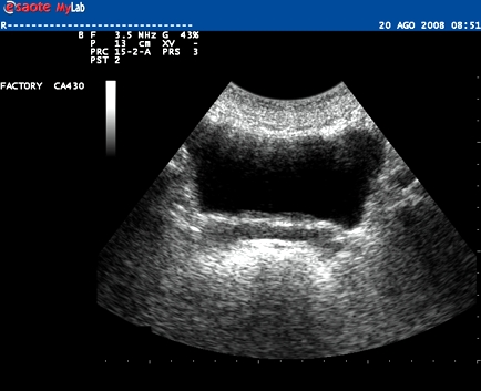
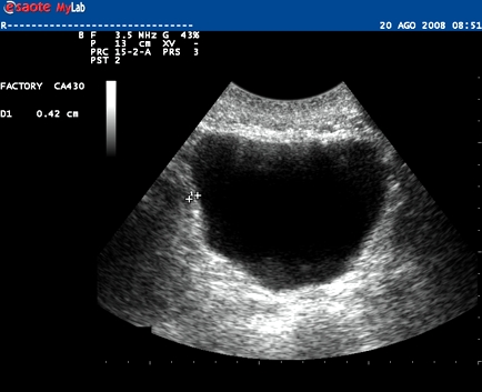
Inclusion date: 13/ 10/2008Ultrasounds made on: 29/05/2008 Instrument: Esaote MegasPatient's age: F 38 years old The patient undergoes an ultrasound examination following an episode of gross hematuria. The images and the video show a hypoechoic formation, which is located in the proximity of the opening of the left ureter and which has a dimension of 1.5xl cm. The formation can be linked to a vesical papilloma which was documented by a cystoscopy. In collaboration with: Dr.ssa Giuliana Santoni, Dr. Pietro Vitali - Jesi (AN) - Italy Presentation: Dr. Massimo Dolciotti - Ancona - Italy Digital processing: Andrea Dini - Ancona - Italy English translation: Prof.ssa Federica Sturani - England
|
|
|
|
|
|
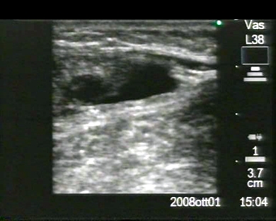
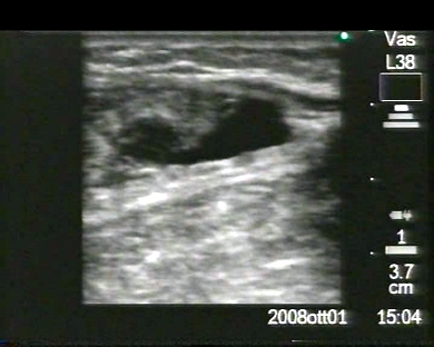
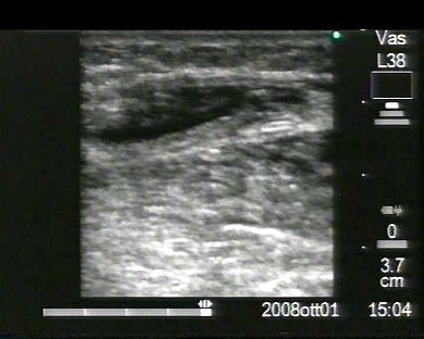
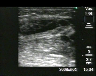
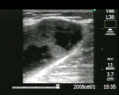
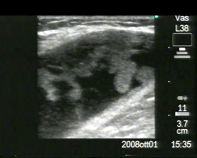
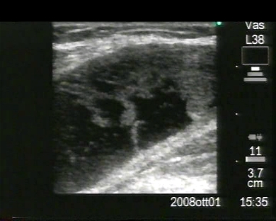
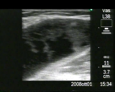
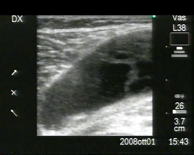
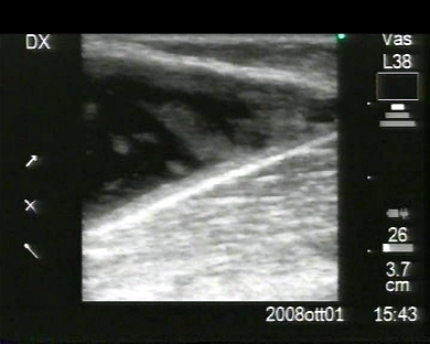
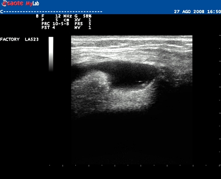
Inclusion Date: 03/ 10/2008Ultrasounds made on: 02/10/2008 Instrument: SonoSite M-TurboPatient's age: F 70 years old The patient undergoes an ultrasound examination following a painless swelling to the right breast. The images show an oval hypo-anechoic formation, with regular and clean margins. The ultrasound structure is uncertain, is it solid or liquid? What is your evaluation? For the discussion we invite you to join the blog in the ecomiei website. Presentation: Dr. Massimo Dolciotti - Ancona - Italy Digital processing: Andrea Dini - Ancona - Italy English translation: Prof.ssa Federica Sturani - England
|
|
|
|
|
|
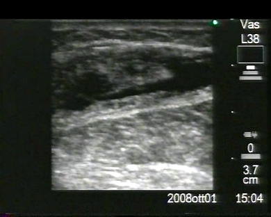
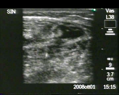
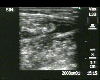
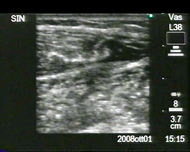
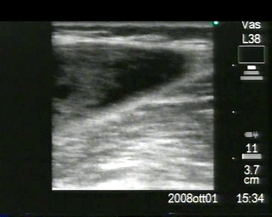
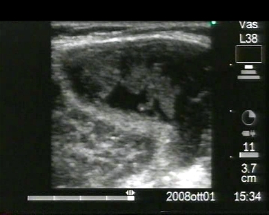
Inclusion Date: 01/ 10/2008Ultrasounds made on: 10/09/2008 Instrument: Esaote MyLab 25Patient's age: M 17 years old The patient undergoes the ultrasound examination following a slightly painful swelling in the right iliac fossa, resulting from a road accident with a motorbike, that happened two months earlier. The images and the video show muscular injury of the second/third grade in some fascicles of muscles of the external oblique muscle, of the internal oblique muscle and of the transversal muscle. In collaboration with: Dr. Daniele Lenti - Jesi (AN) - Italy Presentation: Dr. Massimo Dolciotti - Ancona - Italy Digital processing: Andrea Dini - Ancona - Italy English translation: Prof.ssa Federica Sturani - England
|
|
|
|
|
|
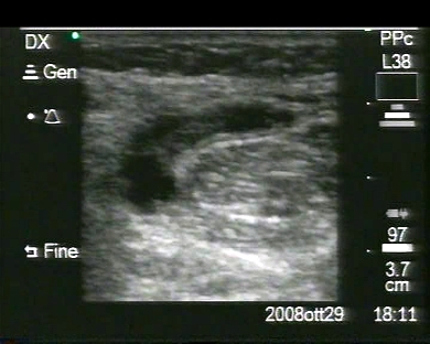
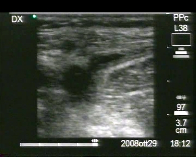
Inclusion Date: 29/ 09/2008Ultrasounds made on: 16/09/2008 Instrument: Toshiba 380APatient's age: M 47 years old The patient undergoes ultrasound examination for a follow-up of an anatomic variation called “dromedary kidney”, usually to be found on the left side and brought about during the foetal period by the compression of the spleen against the third superior of the kidney, which generates a hump on the third medium inferior of the left kidney. Presentation: Dr. Massimo Dolciotti - Ancona - Italy Digital processing: Andrea Dini - Ancona - Italy English translation: Prof.ssa Federica Sturani - England
|
|
|
|