|
|
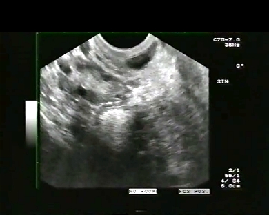
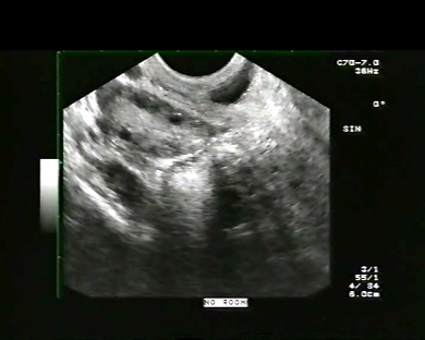
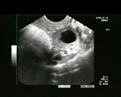
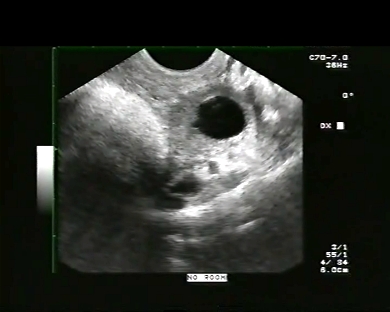
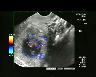
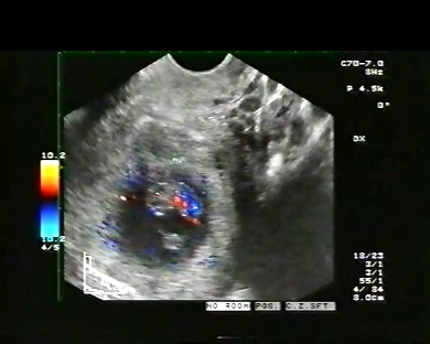
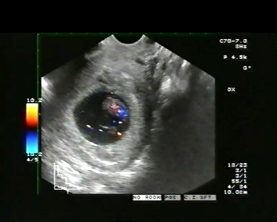
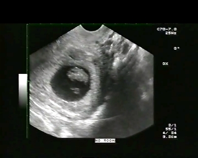
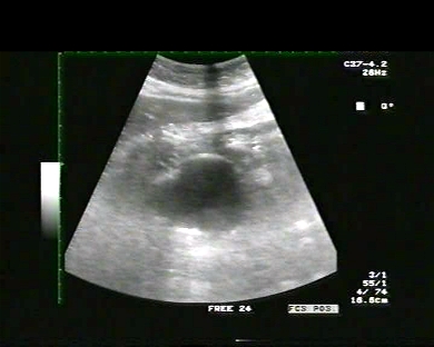
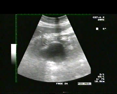
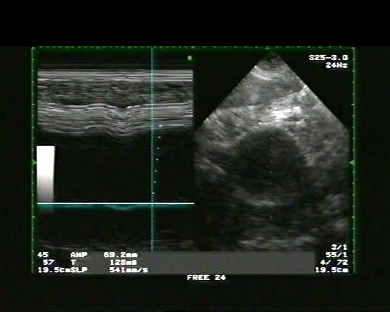
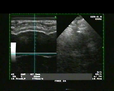
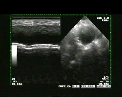
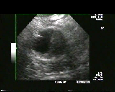
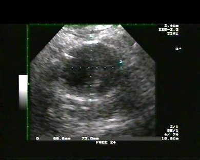
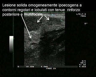
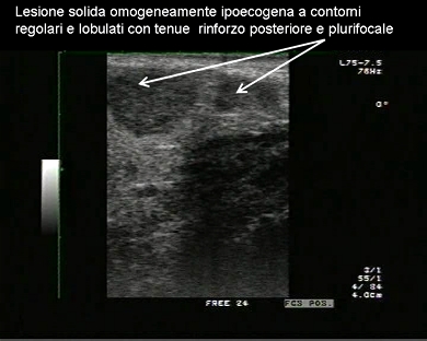
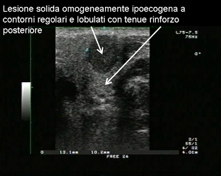
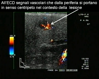
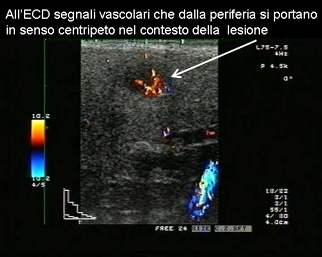
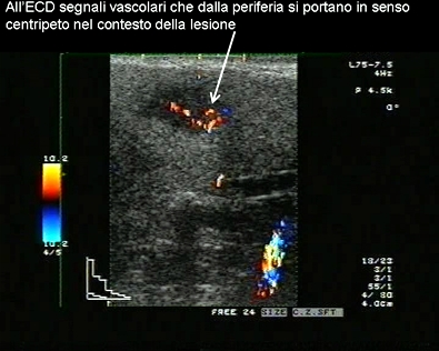
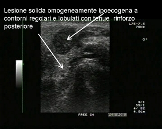
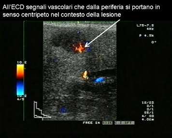
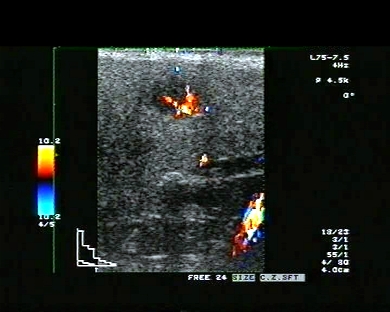
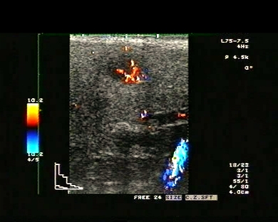
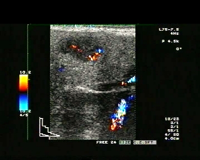
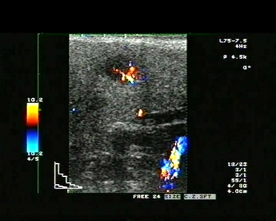
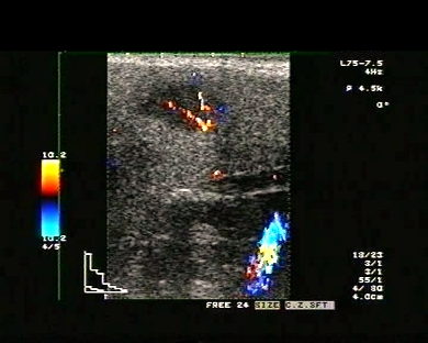
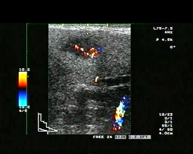
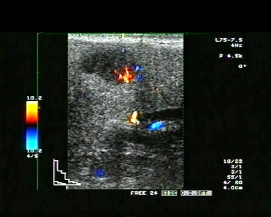
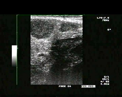
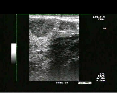
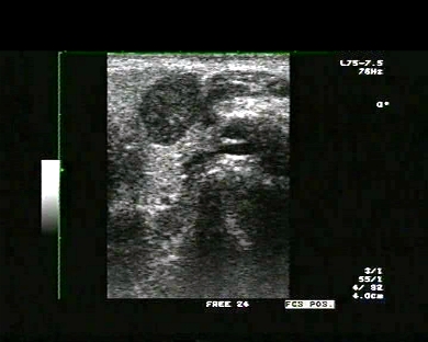
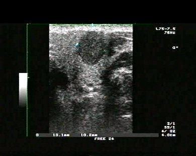
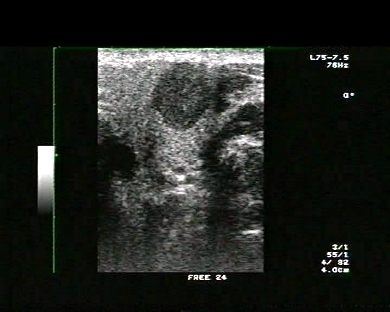
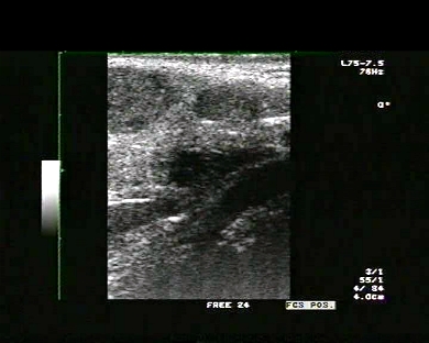
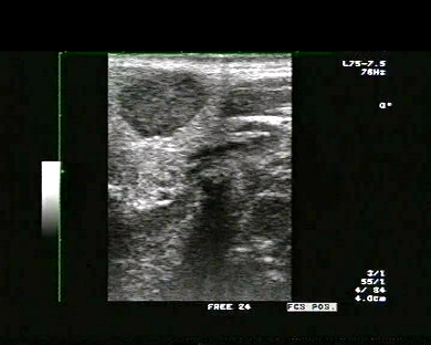
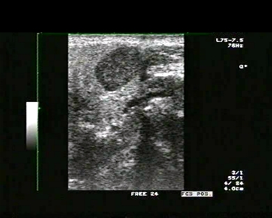
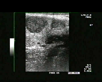
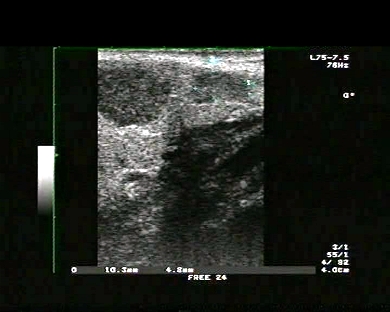
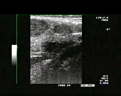
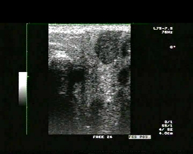
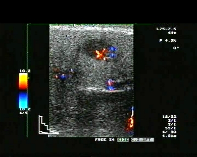
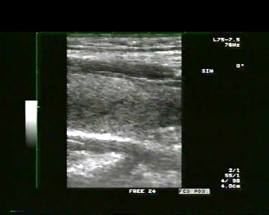
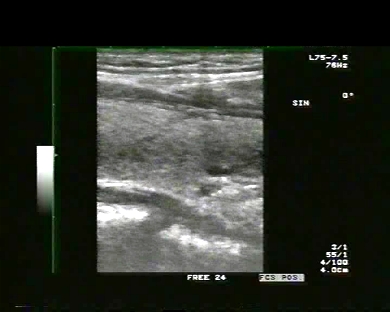
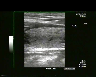
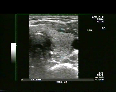
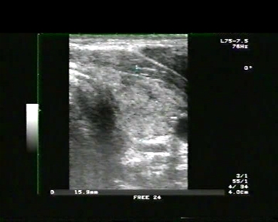
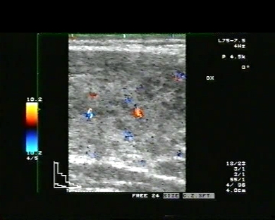
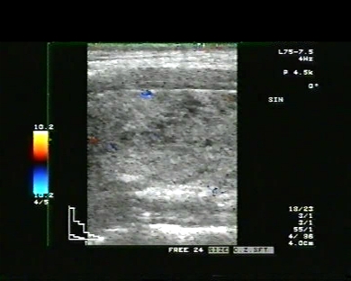
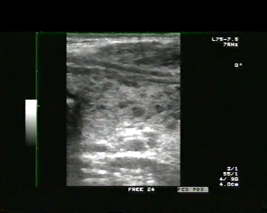
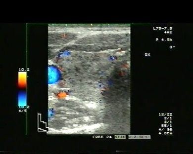
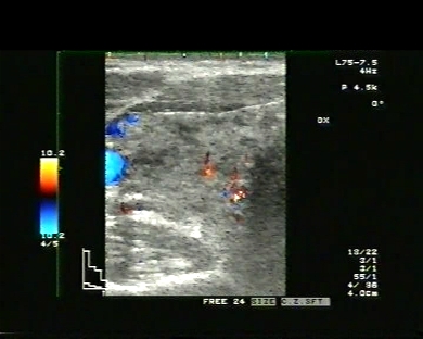
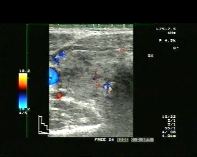
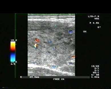
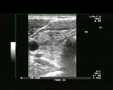
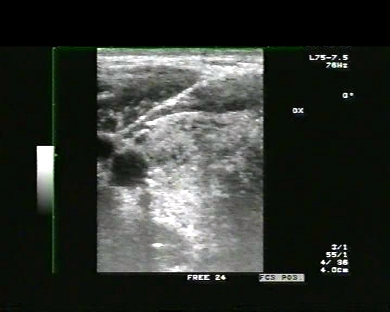
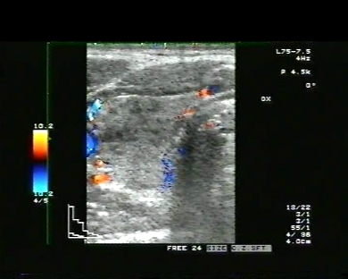
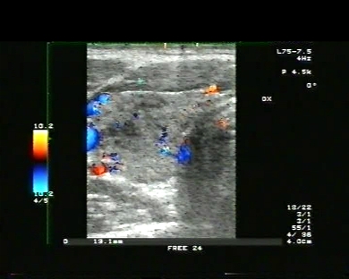
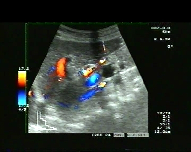
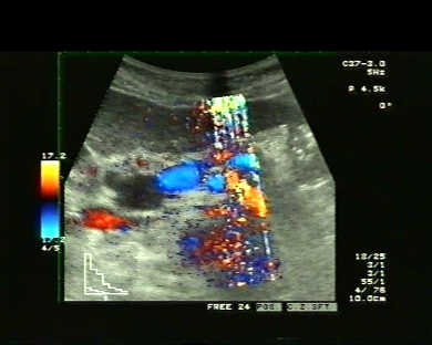
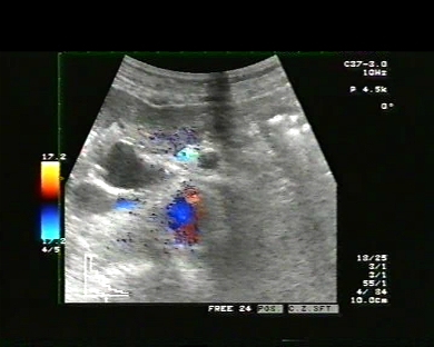
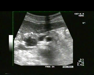
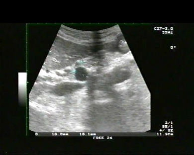
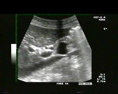
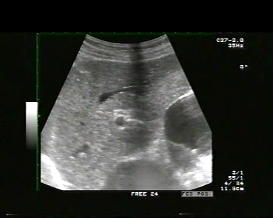
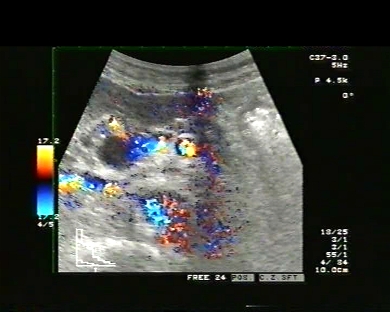
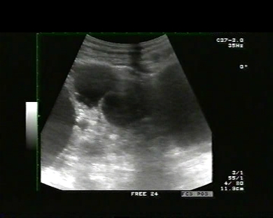
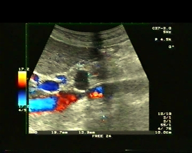
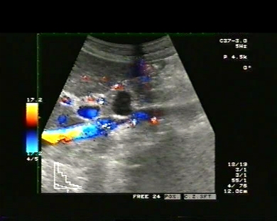
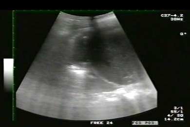
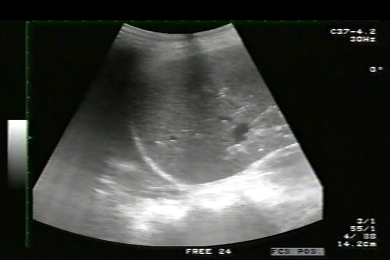
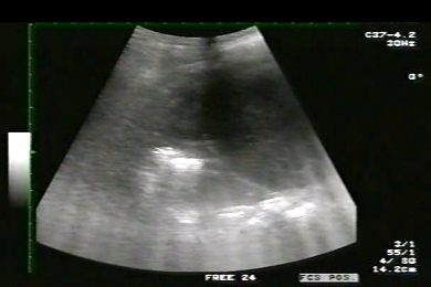
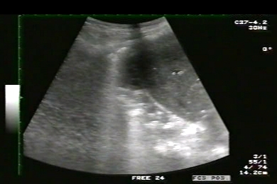
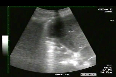
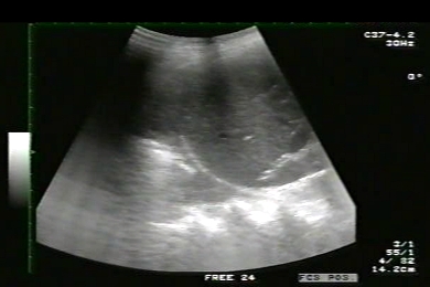
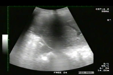
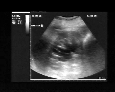
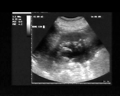
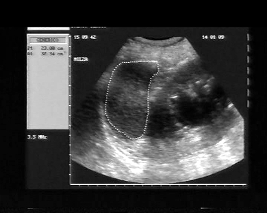
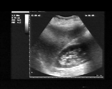
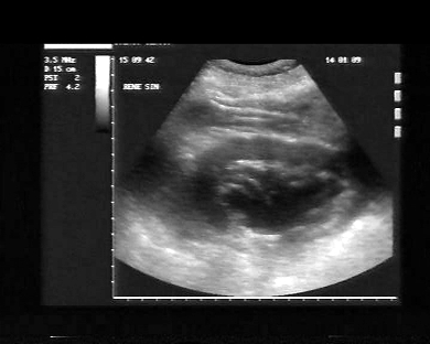
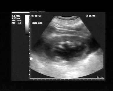
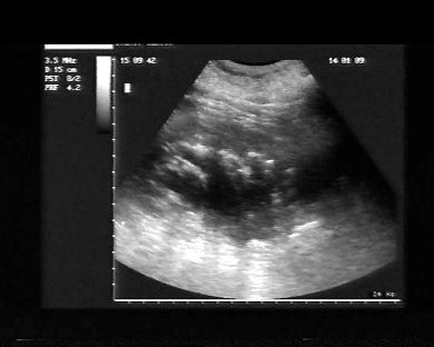
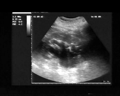
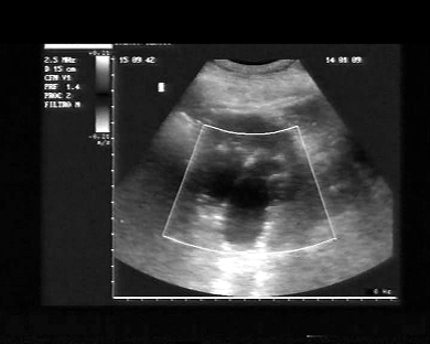
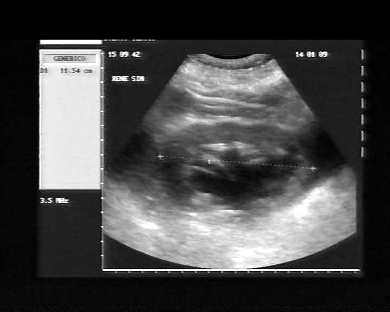
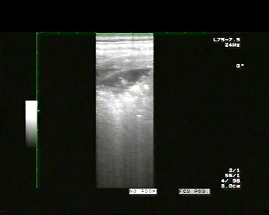
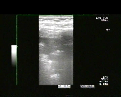
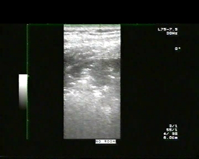
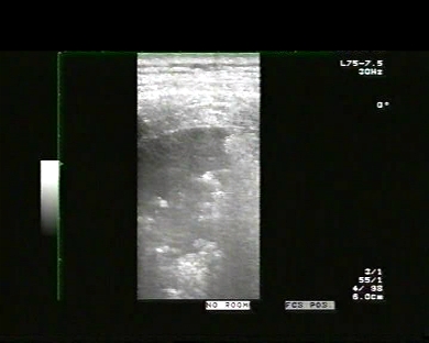
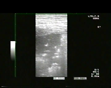
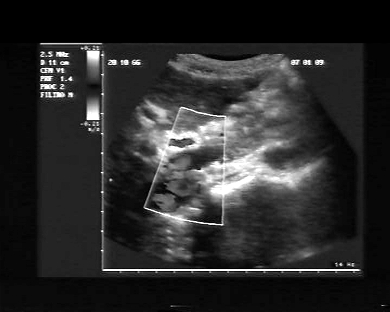
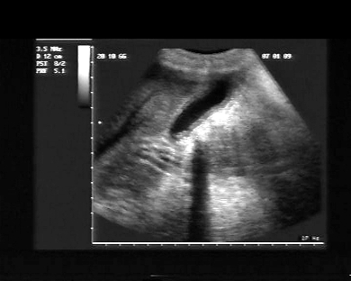
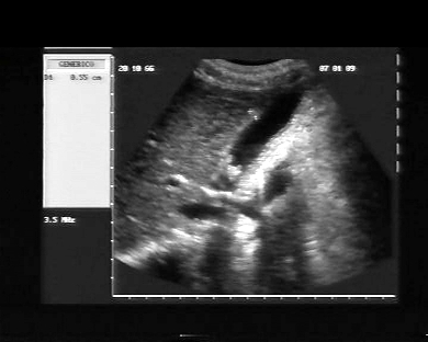
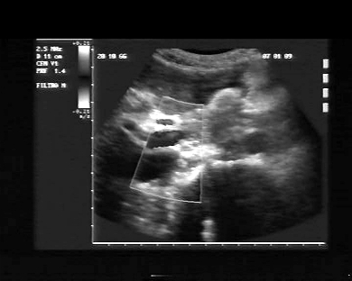
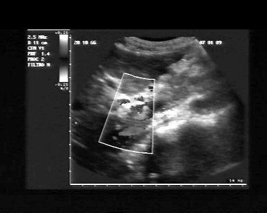
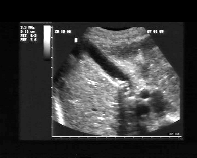
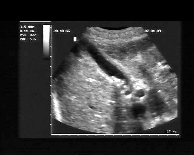
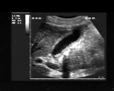
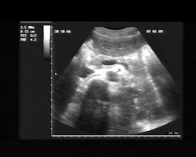
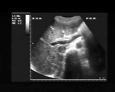
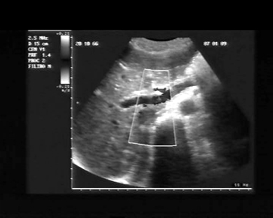
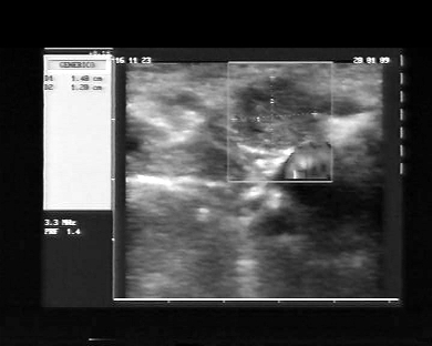
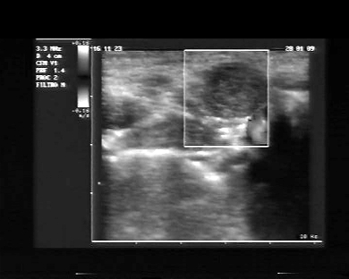
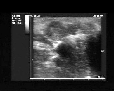
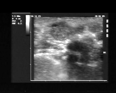
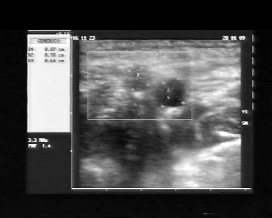
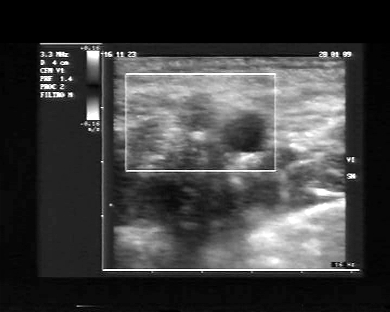
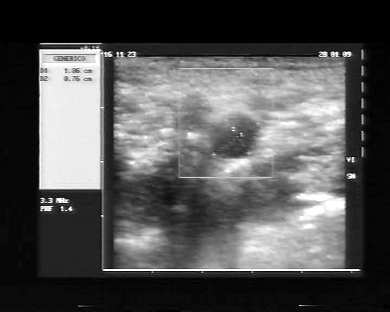
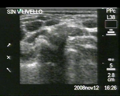
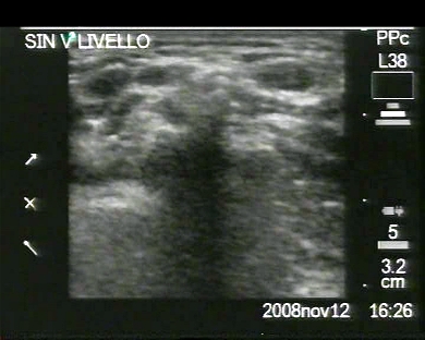
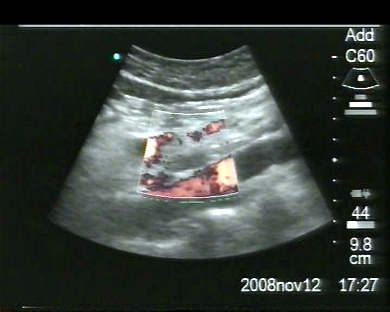
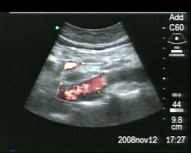
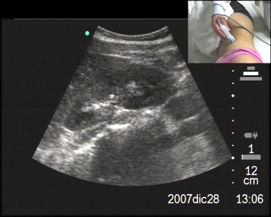
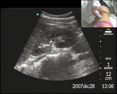
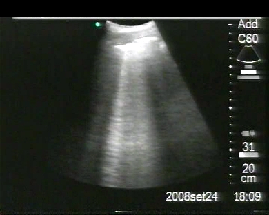
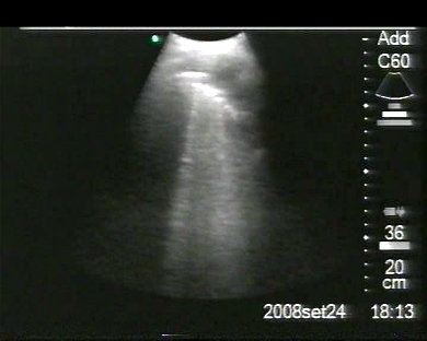
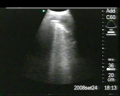
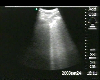
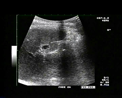
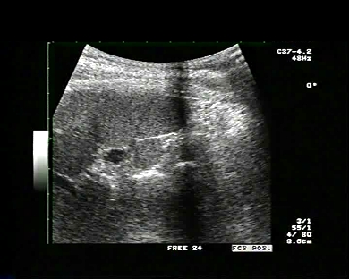
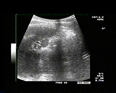
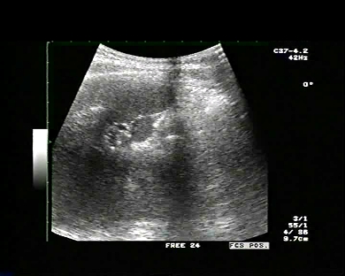
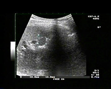
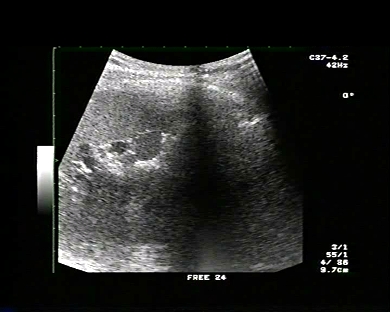
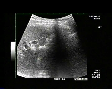
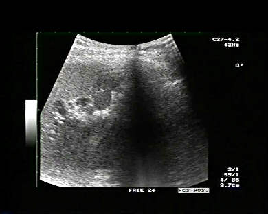
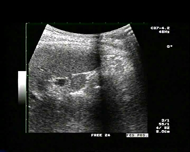
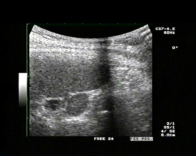
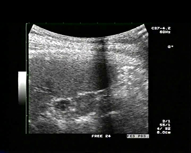
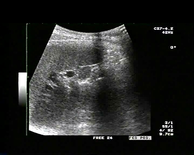
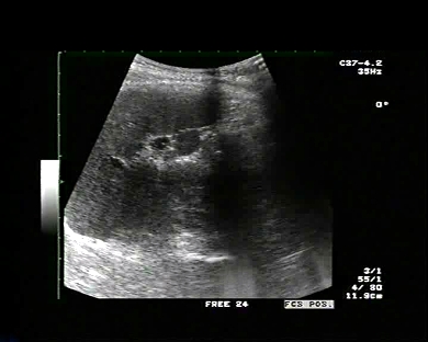
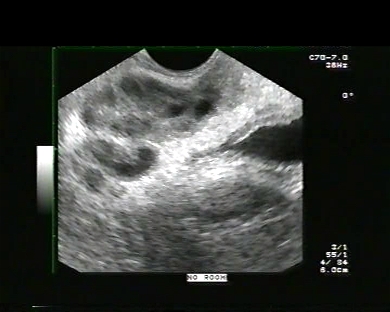
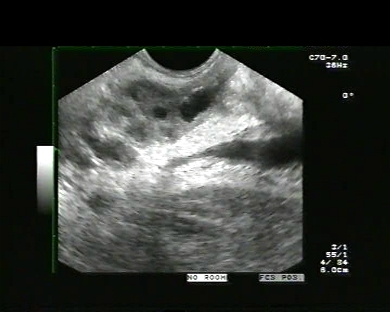
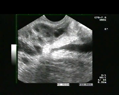
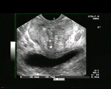
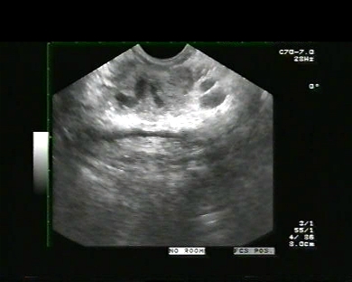
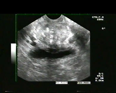
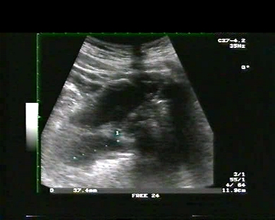
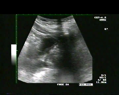
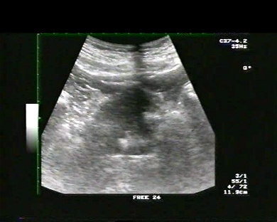
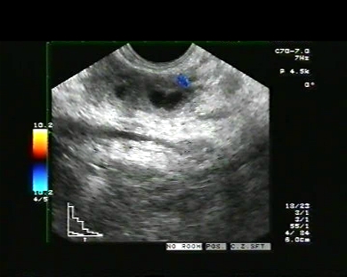
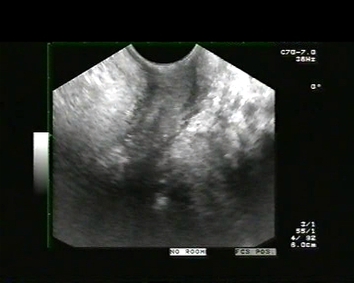
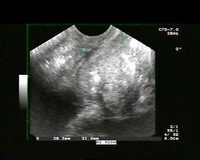
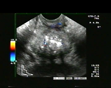
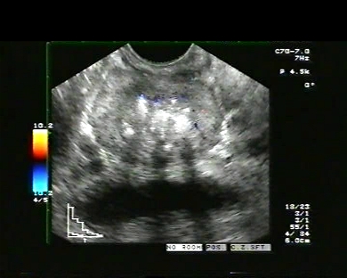
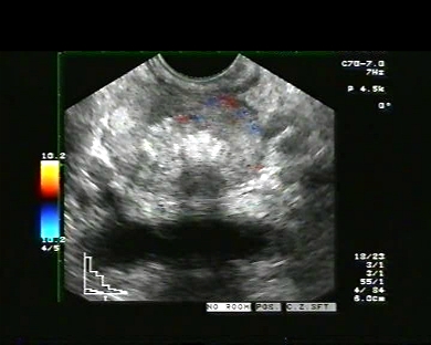
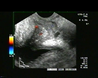
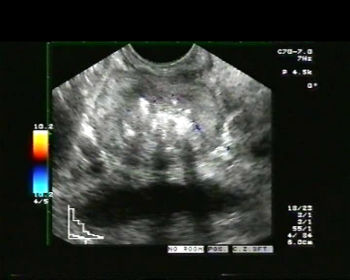
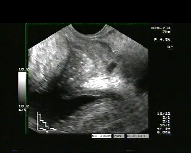
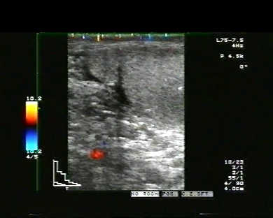
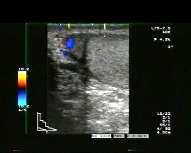
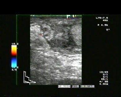
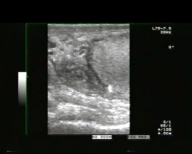
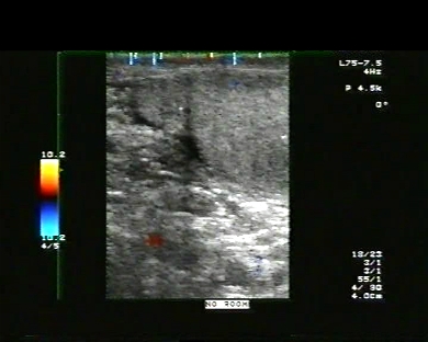
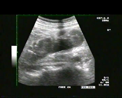
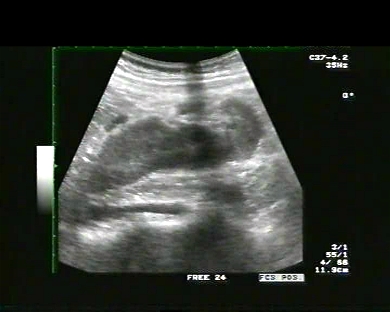
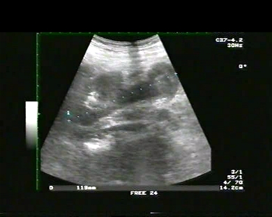
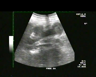
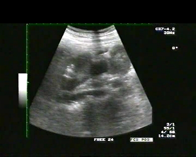
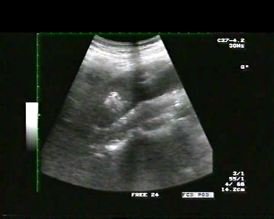
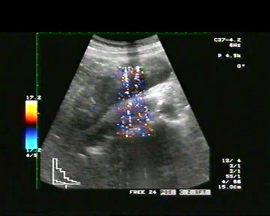
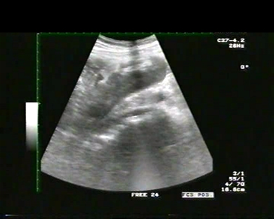
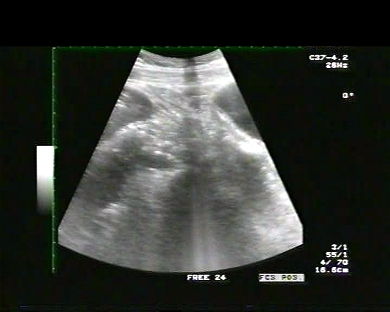
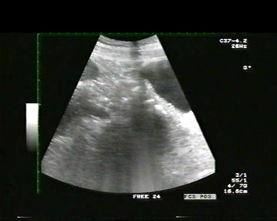
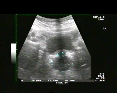
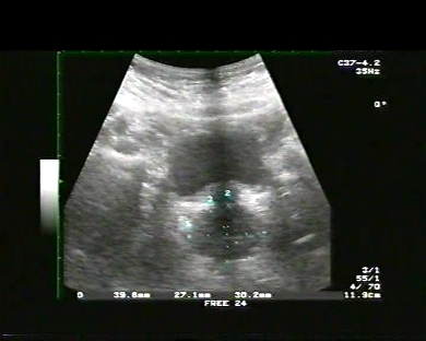
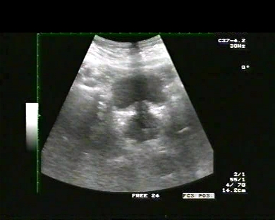
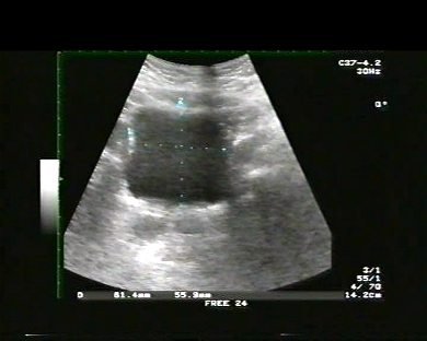
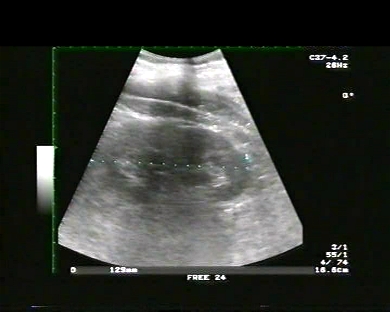
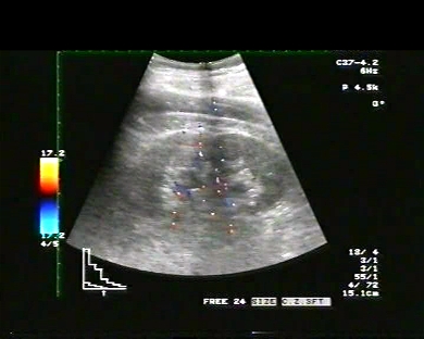
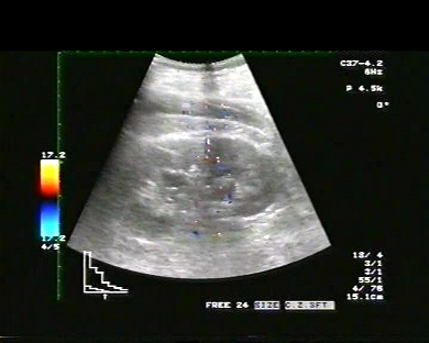
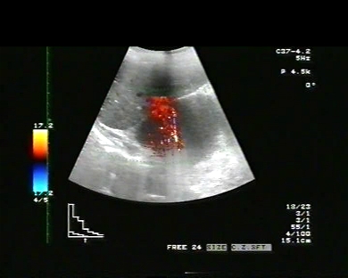
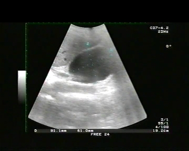
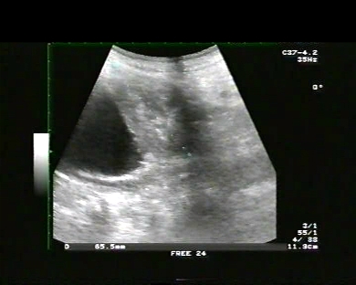
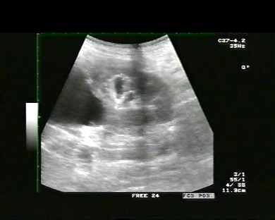
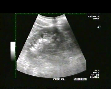
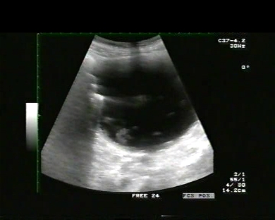
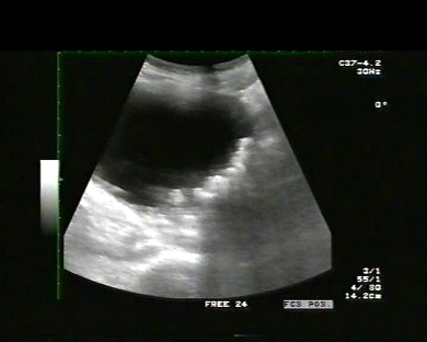
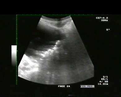
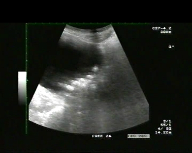
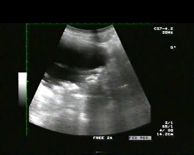
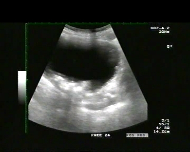
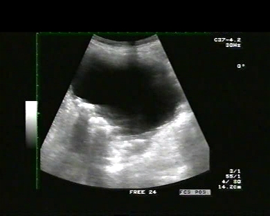
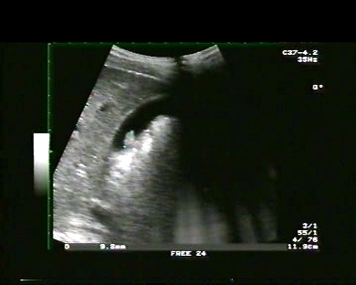
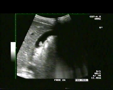
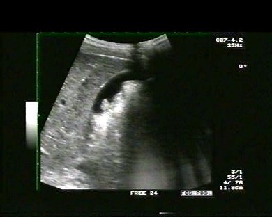
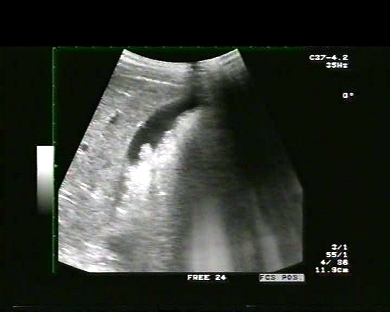
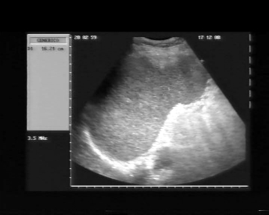
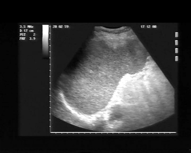
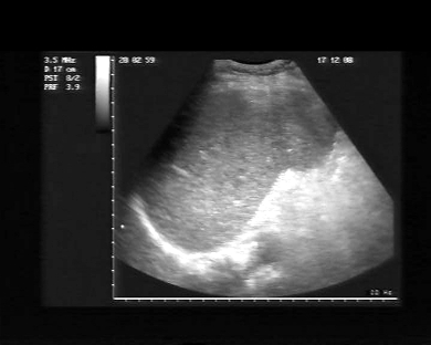
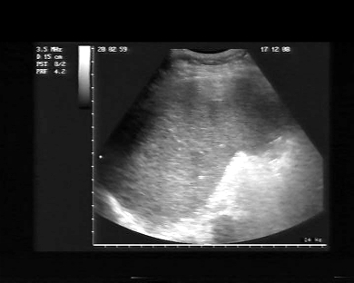
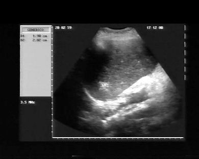
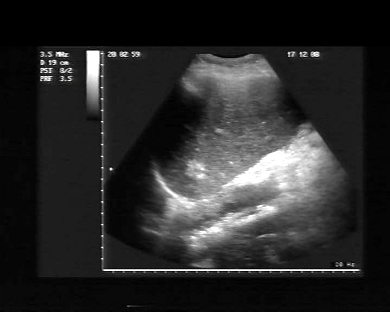
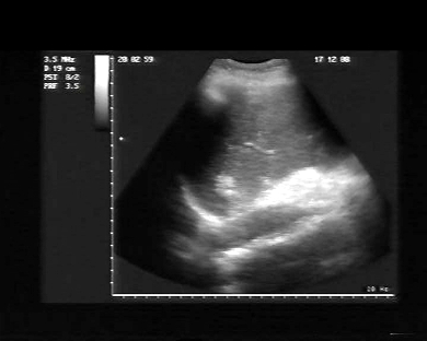
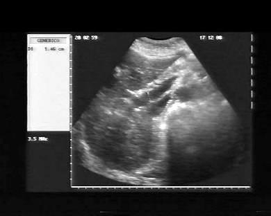
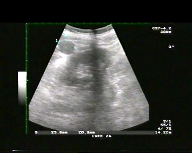
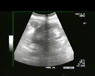
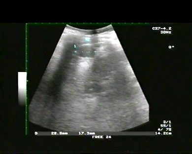
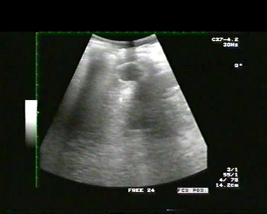
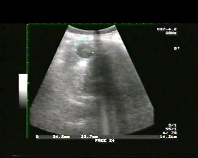
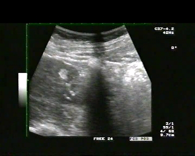
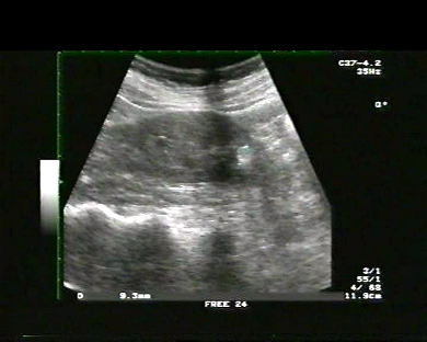
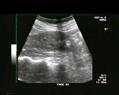
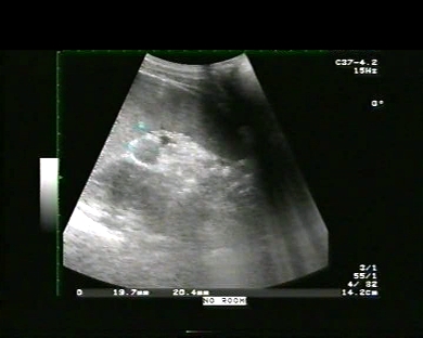
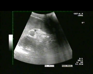
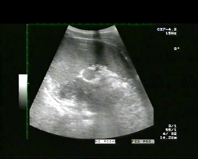
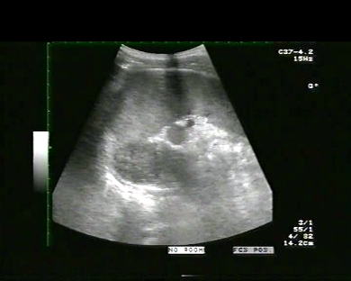
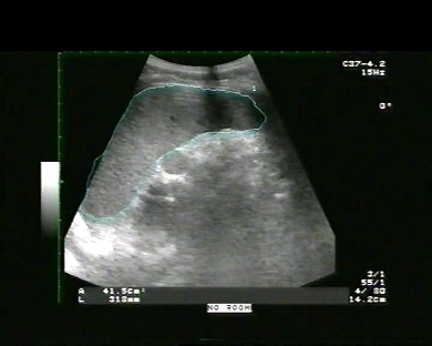
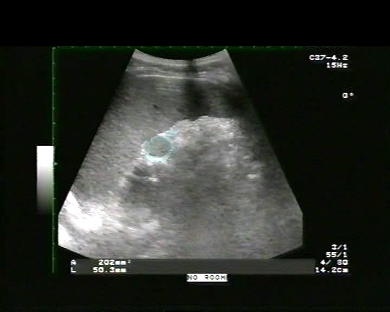
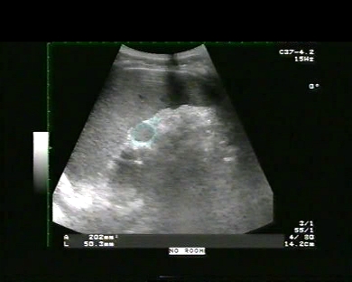
Inclusion date: 30 /03/2009Ultrasound made on: 21/03/2009 Instrument: Toshiba 380A Patient's age: F 76 years old The ultrasound examination is carried out following swelling and pain in the left parotid region. The images and the video show that the left parotid has got some solid and hypoechoic formations, which can be initially explained as a pleomorphic adenoma, or mixed tumor. This is a solid and homogeneously hypoechoic lesion with regular and lobulated borders slightly reinforced at the back. The ECD (Eco-color-Doppler) indicates vascular signals moving within the lesion from the periphery towards the centre. Presentation: Dr. Massimo Dolciotti - Ancona Digital processing: Andrea Dini - Ancona English translation: Prof.ssa Federica Sturani - England
|
|
|
|
|
|
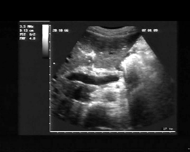
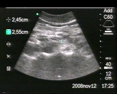
Inclusion date: 25 /03/2009Ultrasound made on: 12/03/2009 Instrument: Toshiba 380A Patient's age: F 20 years old The ultrasound examination is carried out following swelling in the right lateral cervical. The images and the video show the thyroid lobes with a regular echo-structure and morphovolumetry. The right lobe of the thyroid has an anteroposterior diameter of 20mm. The left lobe of the thyroid has got an anteroposterior diameter of 15mm. Presentation: Dr. Massimo Dolciotti - Ancona Digital processing: Andrea Dini - Ancona English translation: Prof.ssa Federica Sturani - England
|
|
|
|
|
|
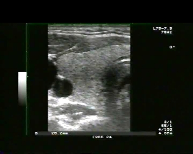
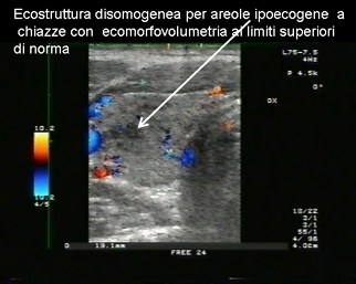
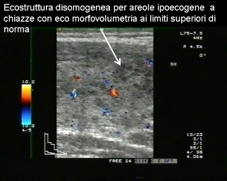
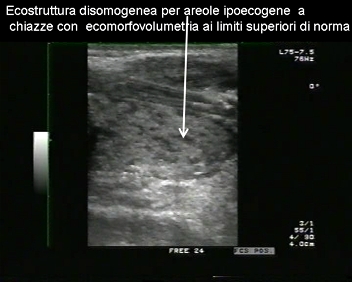
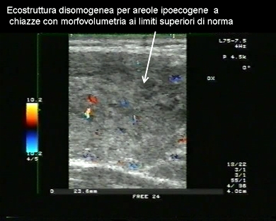
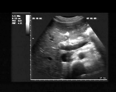
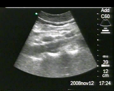
Inclusion date: 23 /03/2009Ultrasound made on: 24/02/2009 Instrument: Toshiba 380A Patient's age: M 20 years old The ultrasound examination is carried out as a laboratory crosscheck of hyperthyroidism (TSH=0.015 mcIU/ml – Normal Range (N.R.) = 0.34 – 5.6 -- Free T3 = 10.40pg/ml – N.R. = 2.20 – 4.20 – Free T4 = 3.87 ng/dl – N.R. = 0.75 – 1.75 – Antireceptor antibodies TSH and anti-Thyroglobulins HTG Normal). The images and the video show the thyroid lobes with a non- homogenous echostructure and hypoechoic areolas, and a reasonable intra-glandular vascularisation. The case history, the lab data and the ultrasound lead towards the autoimmune hyperthyroidism type Hashimoto’s disease. Presentation: Dr. Massimo Dolciotti - Ancona Digital processing: Andrea Dini - Ancona English translation: Prof.ssa Federica Sturani - England
|
|
|
|
|
|
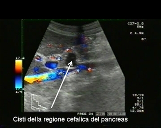
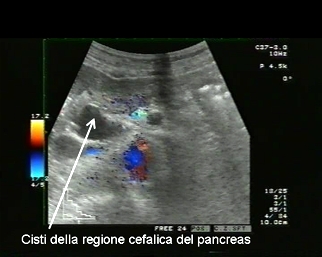
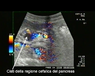
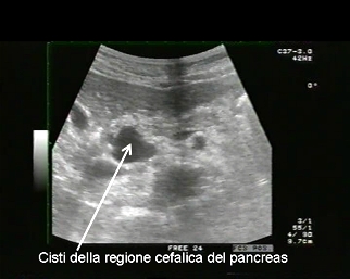
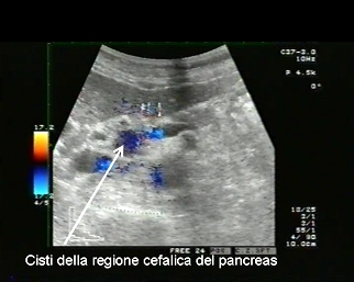
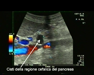
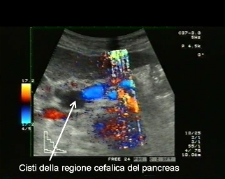
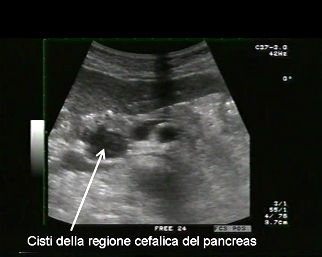
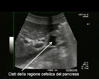
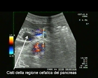
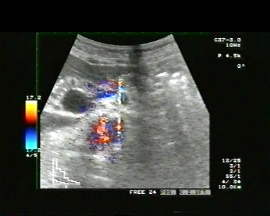
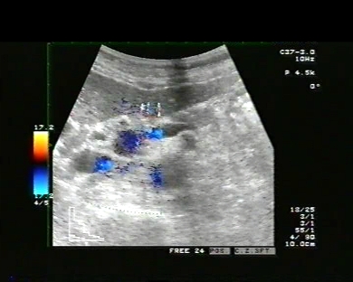
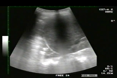
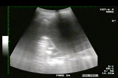
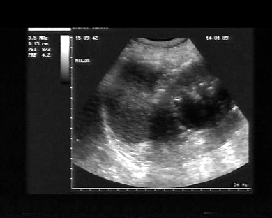
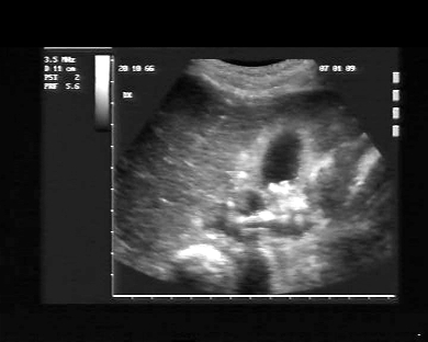
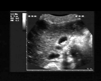
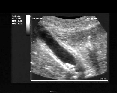
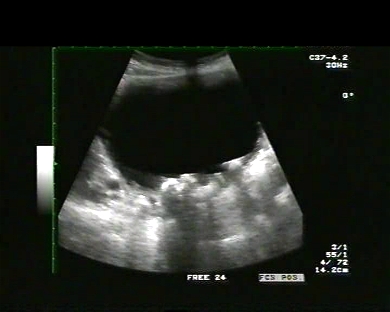
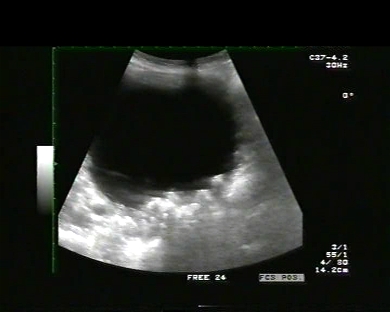
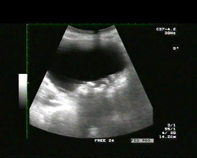
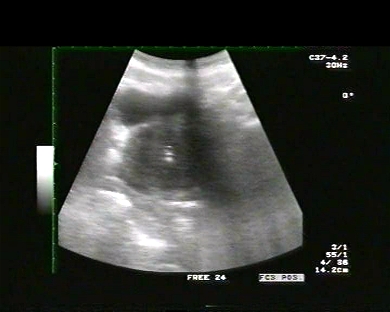
Inclusion date: 18 /03/2009Ultrasound made on: 06/02/2009 Instrument: Toshiba 380A Patient's age: M 86 years old The ultrasound examination to the abdomen is carried out following pain. The images and the video show an anechoic formation with sharp borders located in the cephalic region of the pancreas. The formation can be identified as a liquid cyst of the head of the pancreas in a patient carrying multiple bilateral cysts in the kidneys. In collaboration with: Dr. Quintilio Tomassetti - Ancona Presentation: Dr. Massimo Dolciotti - Ancona Digital processing: Andrea Dini - Ancona English translation: Prof.ssa Federica Sturani - England
|
|
|
|
|
|
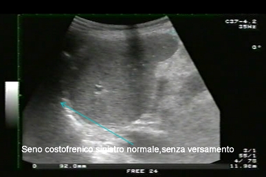
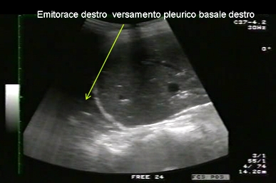
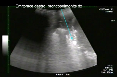
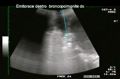
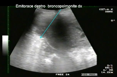
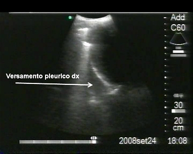
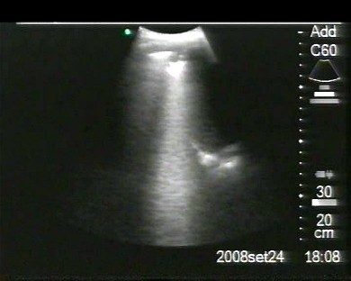
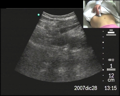
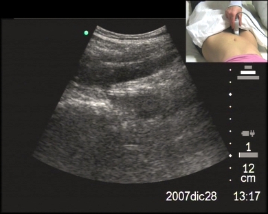
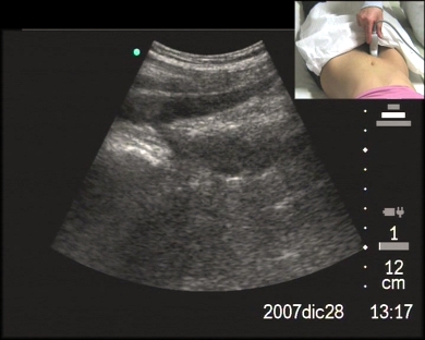
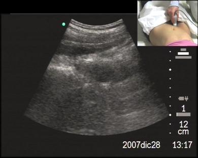
Inclusion date: 11 /03/2009Ultrasound made on: 24/02/2009 Instrument: Toshiba 380A Patient's age: F 68 years old The ultrasound examination in the abdomen and in the thorax is carried out following fever and epigastric pains irradiated to the right hypochondrium. The examination highlights a rise of the right lung base and hypophonesis. The images and the video show a hyperechoic image created by an inflammatory thickening in the lung inferior parenchyma and by a parapneumonnic effusion at the base of the right hemitorax. In collaboration with: Dr. Quintilio Tomassetti - Ancona Presentation: Dr. Massimo Dolciotti - Ancona Digital processing: Andrea Dini - Ancona English translation: Prof.ssa Federica Sturani - England
|
|
|
|
|
|
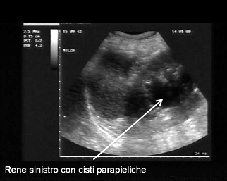
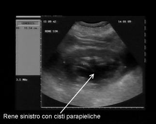
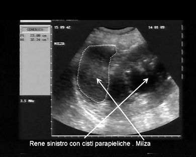
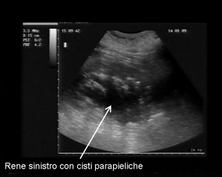
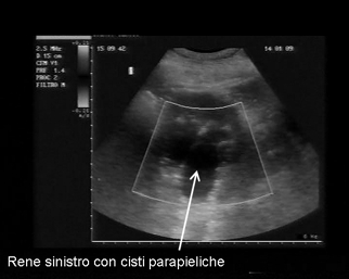
Inclusion date: 02 /03/2009Ultrasound made on: 14/01/2009 Instrument: Esaote Megas Patient's age: M 66 years old The ultrasound examination in the abdomen is carried out as a follow-up to an adeno-carcinoma of the colon. The images and the video show the left kidney in its position and parapyelic cysts. The right kidney is in its position and there are small parapyelic cysts. In collaboration with: Dr. Pietro Vitali - Jesi (AN) Presentation: Dr. Massimo Dolciotti - Ancona Digital processing: Andrea Dini - Ancona English translation: Prof.ssa Federica Sturani - England
|
|
|
|
|
|
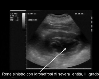
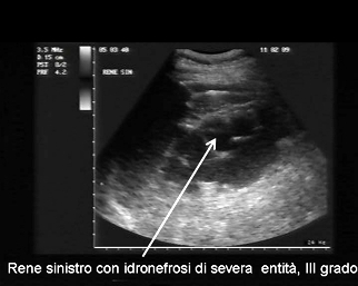
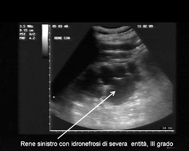
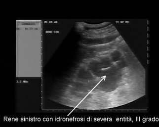
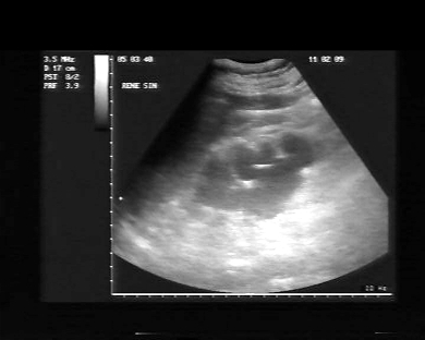
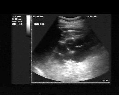
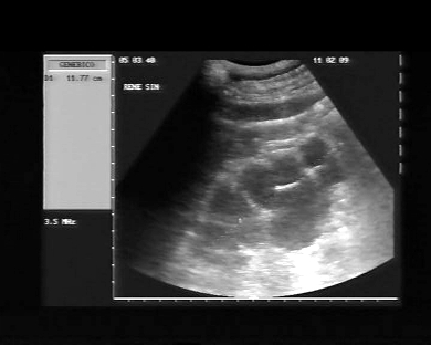
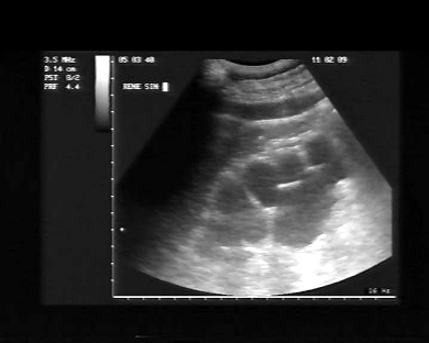
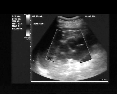
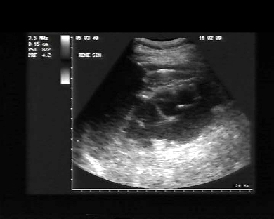
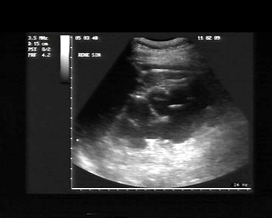
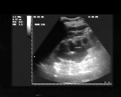
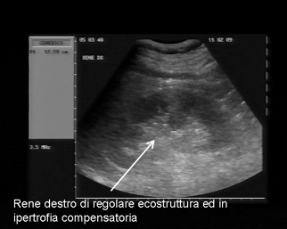
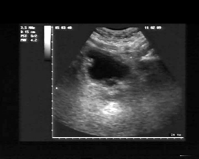
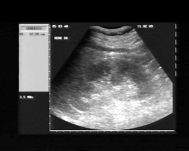
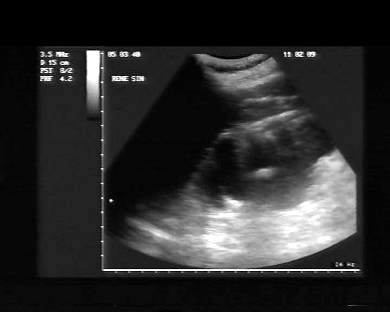
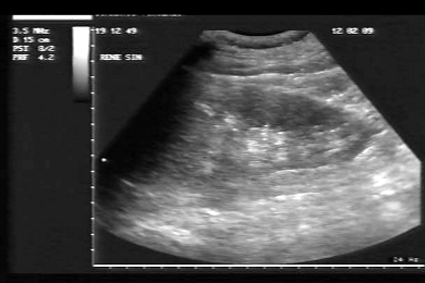
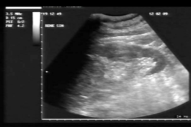
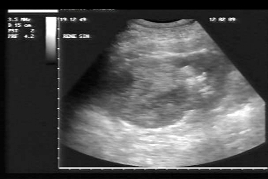
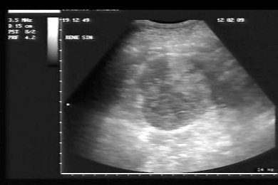
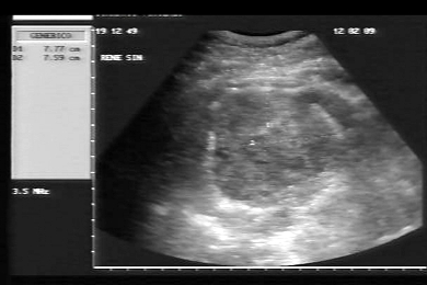
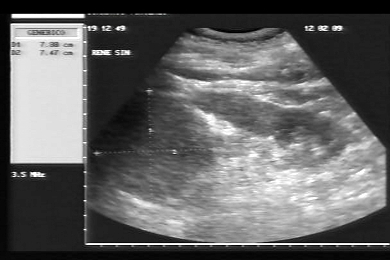
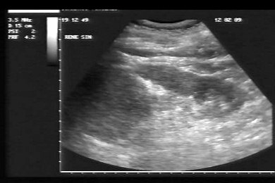
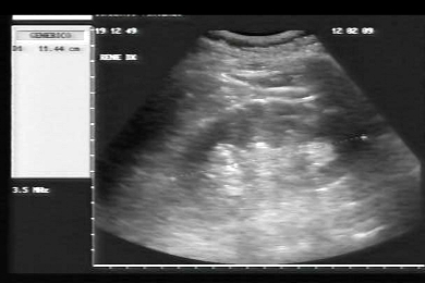
Inclusion date: 02 /03/2009Ultrasound made on: 11/02/2009 Instrument: Esaote Megas Patient's age: M 68 years old The ultrasound examination in the abdomen is carried out following pain in the left hip. The images and the video show the left kidney affected by severe hydronephrosis (III grade), and a reduction of the thickness of the cortical medullar region. Left kidney in position, with regular echostructure and in compensatory hypertrophy. In collaboration with: Dr. Daniele Lenti - Jesi (AN) Presentation: Dr. Massimo Dolciotti - Ancona Digital processing: Andrea Dini - Ancona English translation: Prof.ssa Federica Sturani - England
|
|
|
|
|
|
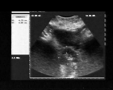
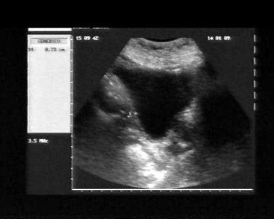
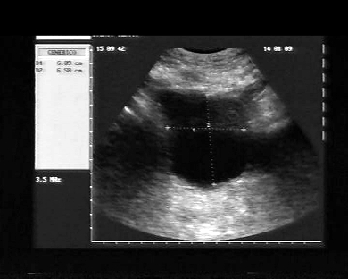
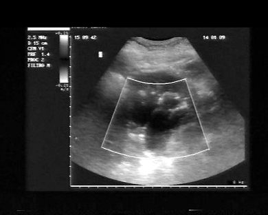
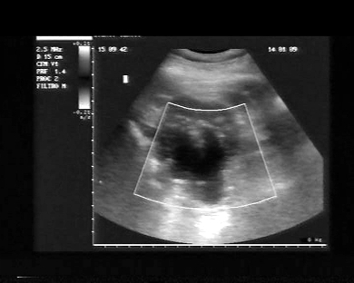
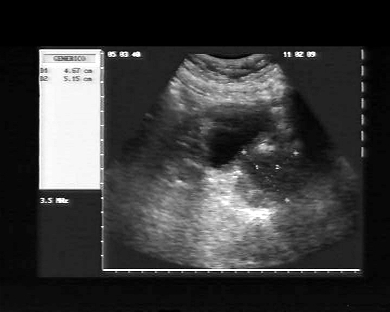
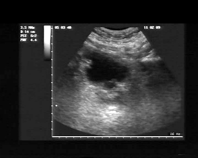
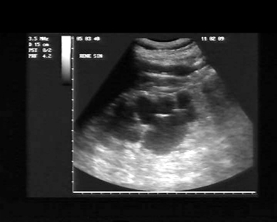
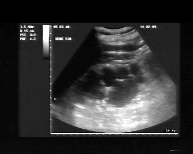
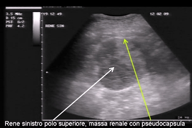
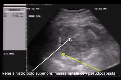
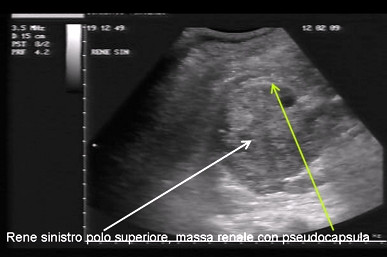
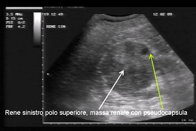
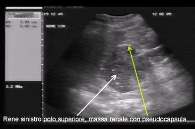
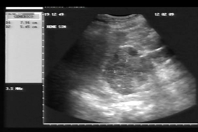
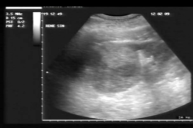
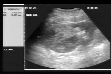
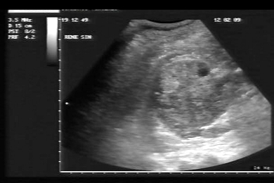
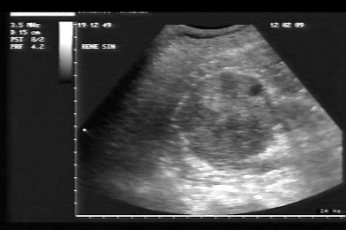
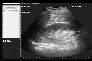
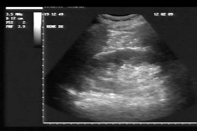
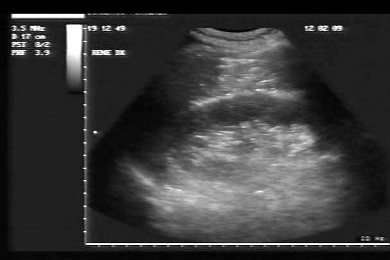
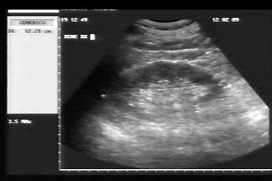
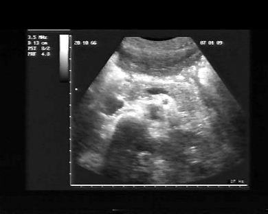
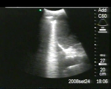
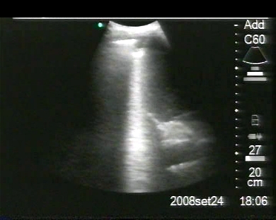
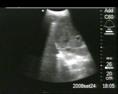
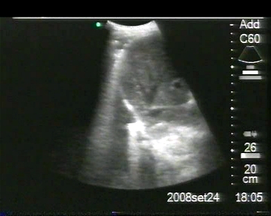
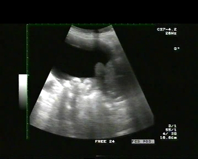
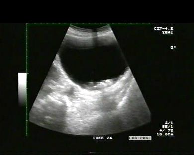
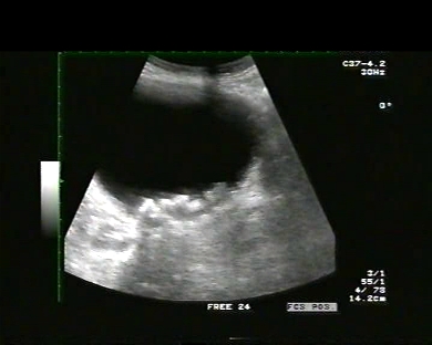
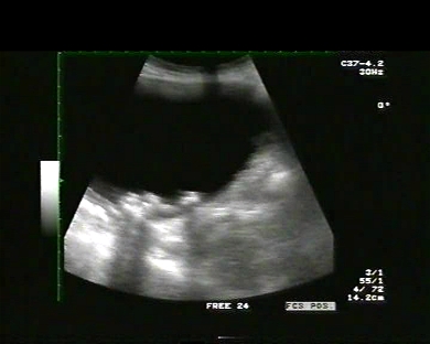
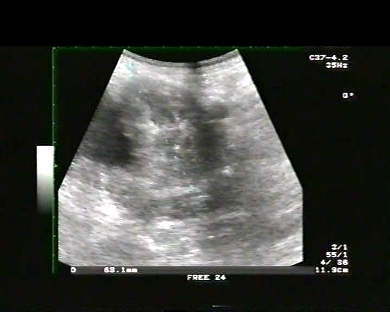
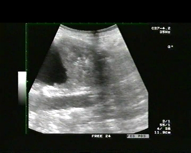
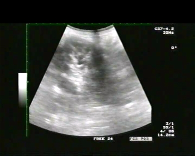
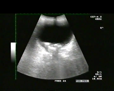
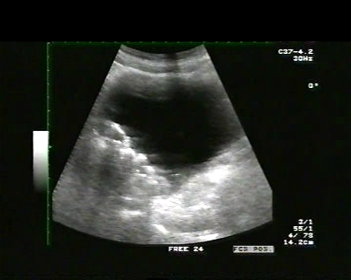
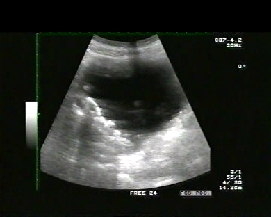
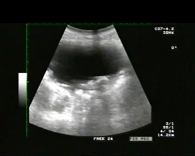
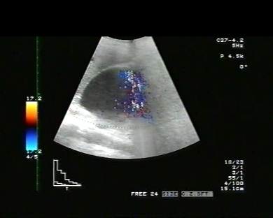
Inclusion date: 27 /02/2009Ultrasound made on: 12/02/2009 Instrument: Esaote Megas Patient's age: M 59 years old The ultrasound examination in the abdomen is carried out following the appearance three days earlier of gross hematuria not accompanied by any other symptoms. The images and the video show a solid mass in the superior pole of the left kidney, which alters the kidney profile. The mass has a dimension of 79x89 mm, a non-homogeneous echostructure and is delimited by a pseudo-capsule with intralesional vascularisation. The right kidney is in position and with a regular echostructure and morphovolumetry. The bladder is scarcely full and appears with regular walls and without endoluminal images. The prostate has a regular echostructure and morphovolumentry. Esame istologico: neoplasia renale in sede polare superiore di 6,5 cm di diametro, di colore giallastro, con aree emorragiche. Carcinoma renale a cellule chiare grado 2 secondo Fuhrman. La neoplasia infiltra la capsula ed il grasso perirenale. Surrene sinistro indenne. In collaboration with: Dr. Pietro Vitali - Jesi (AN) Presentation: Dr. Massimo Dolciotti - Ancona Digital processing: Andrea Dini - Ancona English translation: Prof.ssa Federica Sturani - England
|
|
|
|
|
|
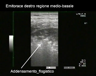
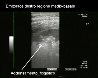
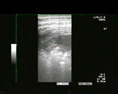
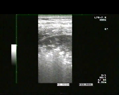
Inclusion date: 18 /02/2009Ultrasound made on: 23/01/2009 Instrument: Toshiba 380A Patient's age: M 51 years old The ultrasound examination of the chest is carried out following a cough, which started a few days earlier, and fever (38° C) which appeared on the same day of the examination. For this examination both a convex and a linear probe has been used. The images and the video show inflammatory clotting in the right middle-basal area, without pleural effusion. In collaboration with: Dr. Quintilio Tomassetti - Ancona Presentation: Dr. Massimo Dolciotti - Ancona Digital processing: Andrea Dini - Ancona English translation: Prof.ssa Federica Sturani - England
|
|
|
|
|
|
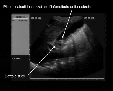
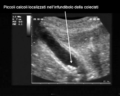
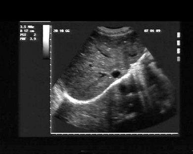
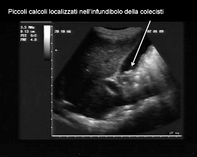
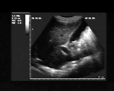
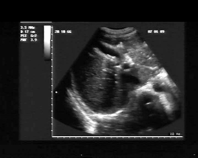
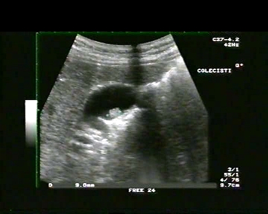
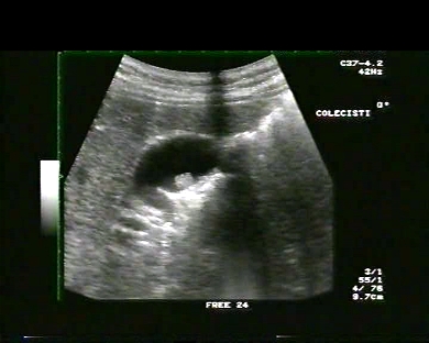
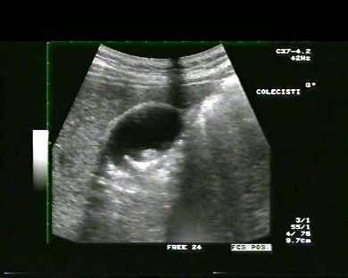
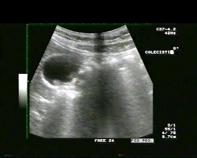
Inclusion date: 13 /02/2009Ultrasound made on: 07/01/2009 Instrument: Esaote Megas Patient's age: F 42 years old The ultrasound examination is carried out following abdominal colic which started three days before. The images and the video show the cholecyst with small stones inside located in the infundibulum. The wall of the cholecyst is thickened, the examination show the cystic duct, the common bile duct does not show dilations and the intrahepatic biliary tract is also normal. In collaboration with: Dr. Daniele Lenti - Jesi (AN) Presentation: Dr. Massimo Dolciotti - Ancona Digital processing: Andrea Dini - Ancona English translation: Prof.ssa Federica Sturani - England
|
|
|
|
|
|
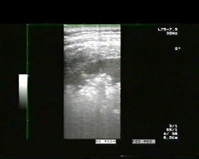
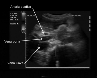
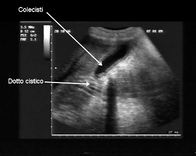
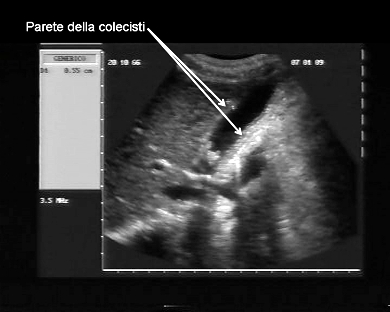
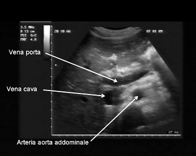
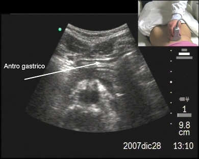
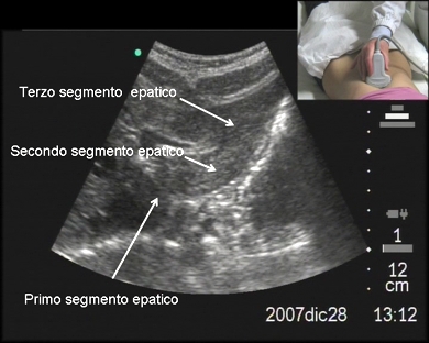
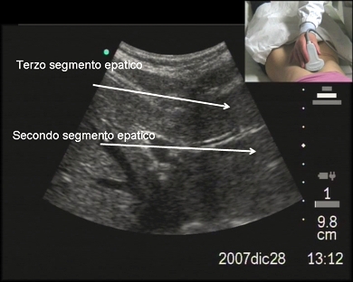
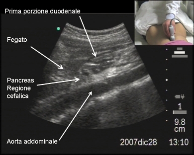
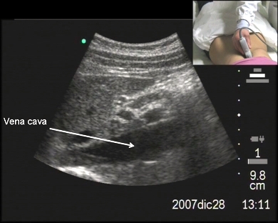
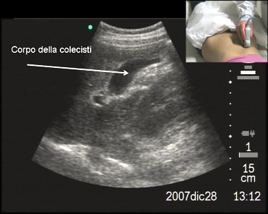
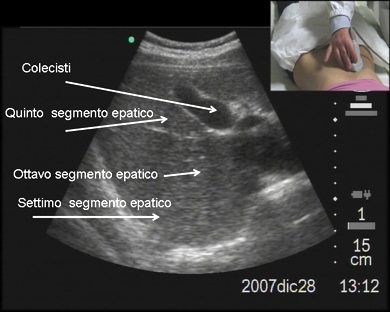
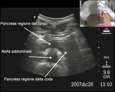
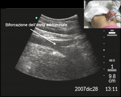
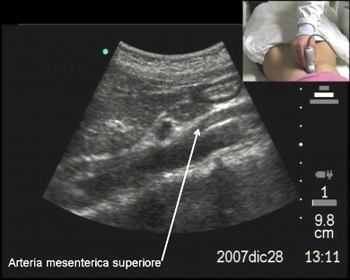
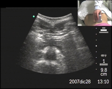
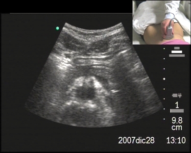
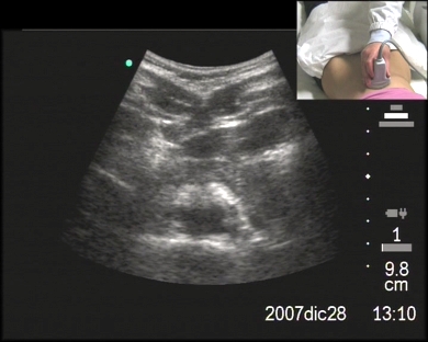
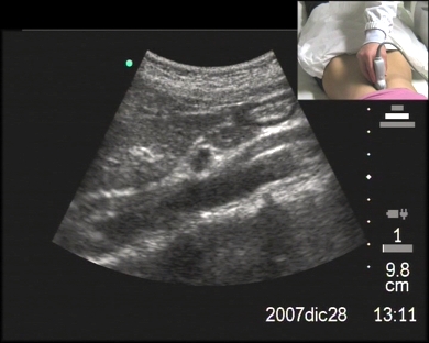
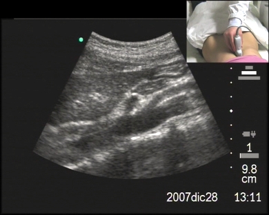
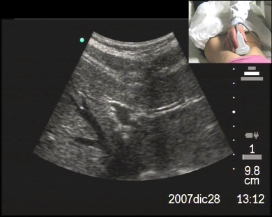
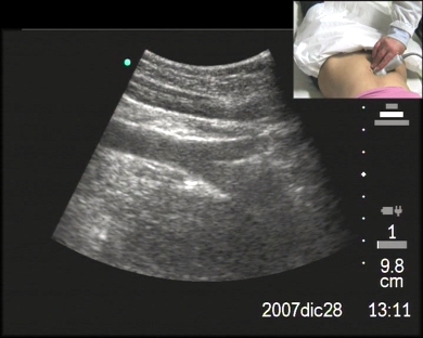
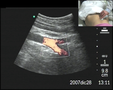
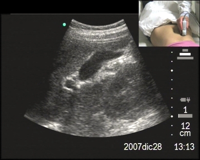
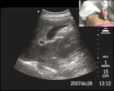
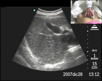
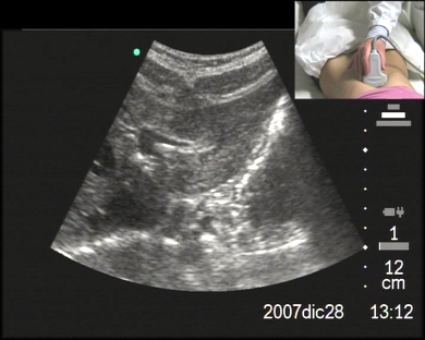
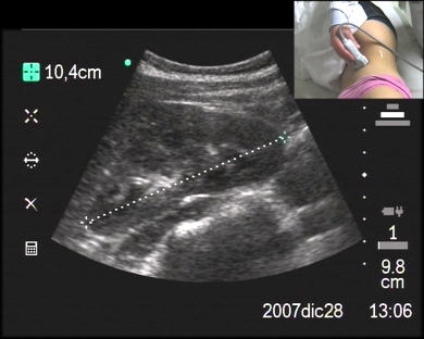
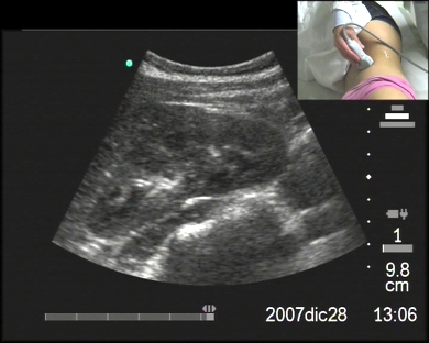
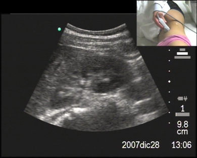
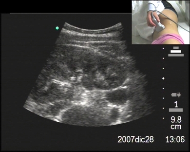
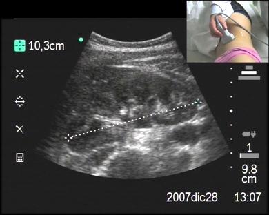
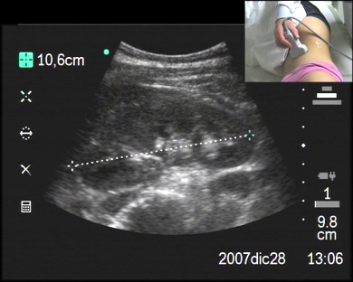
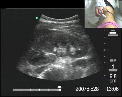
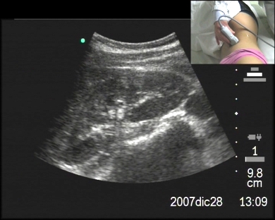
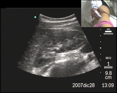
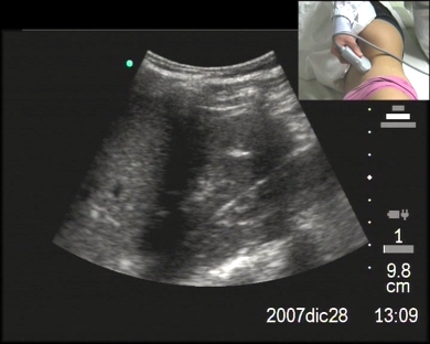
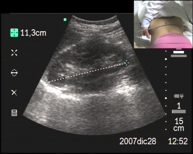
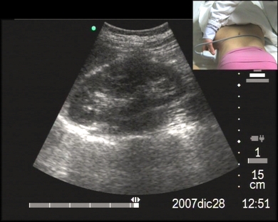
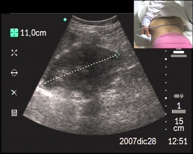
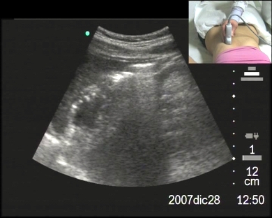
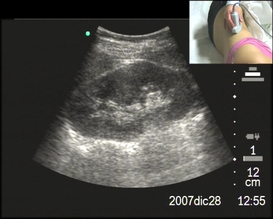
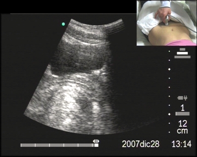
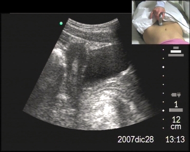
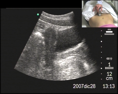
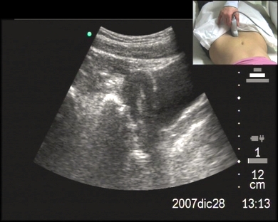
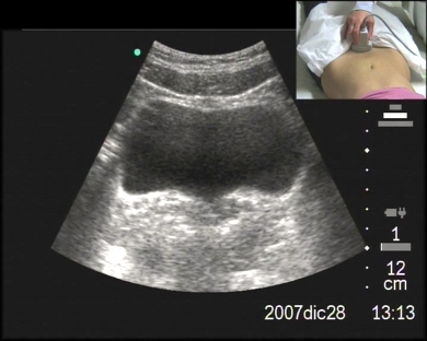
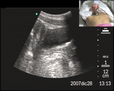
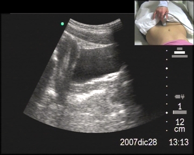
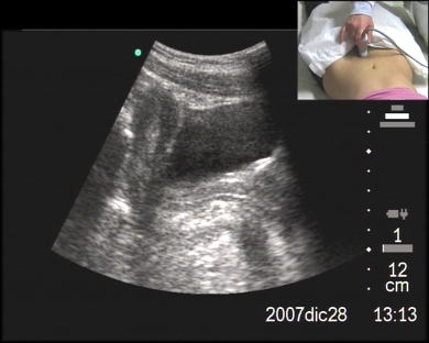
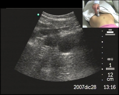
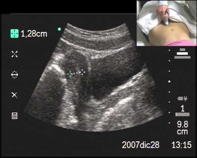
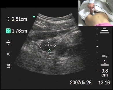
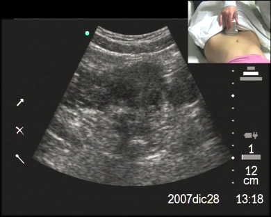
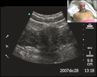
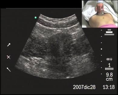
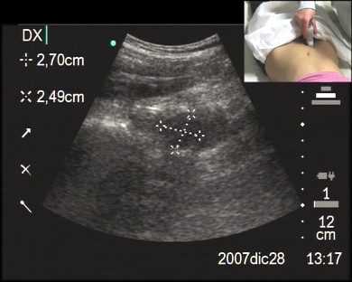
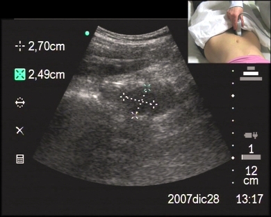
Inclusion date: 11 /02/2009Ultrasound made on: 28/12/2007 Instrument: Sonosite Patient's age: F 18 years old The ultrasound examination is carried out to study the upper abdomen. The images and the video show the liver with hepatic segments, the cholecyst, the pancreas, the abdominal aorta, the vena cava, the superior mesenteric artery and the gastric antrum. Presentation: Dr. Massimo Dolciotti - Ancona Digital processing: Andrea Dini - Ancona English translation: Prof.ssa Federica Sturani - England
|
|
|
|
|
|
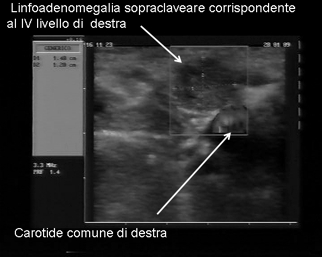
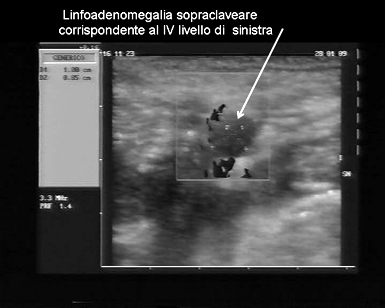
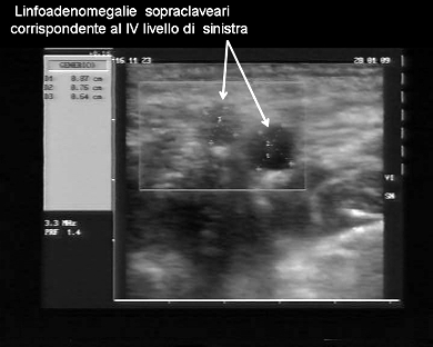
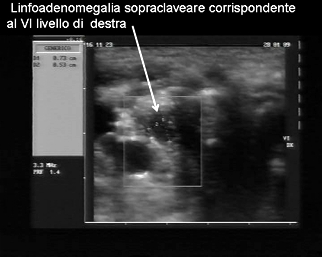
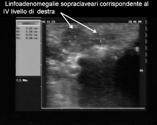
Inclusion date: 11 /02/2009Ultrasound made on: 20/01/2009 Instrument: Esaote Megas Patient's age: F 85 years old The ultrasound examination is carried out as a follow-up to a papillary carcinoma of the thyroid in a patient who had already undergone a thyroidectomy. The images and the video show lympho-adenomegalies in the right and left supraclavicular and retrosternal areas, which are likely to house metastates. In collaboration with: Dr. Pietro Vitali (oncologist–surgeon) - Jesi (AN) Presentation: Dr. Massimo Dolciotti - Ancona Digital processing: Andrea Dini - Ancona English translation: Prof.ssa Federica Sturani - England
|
|
|
|
|
|
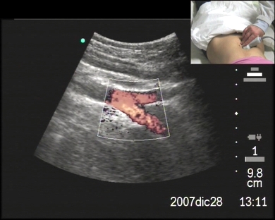
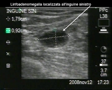
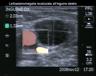
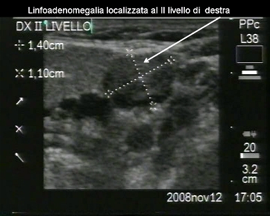
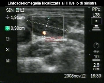
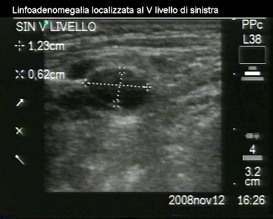
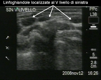
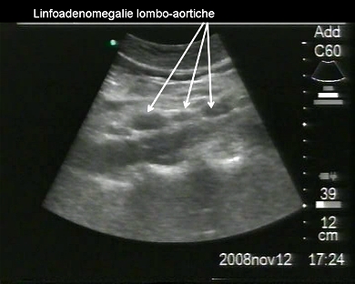
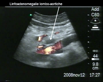
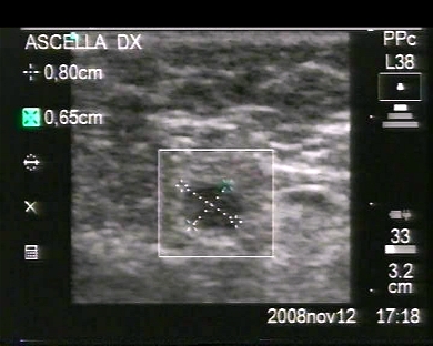
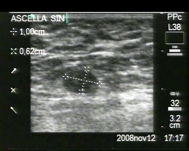
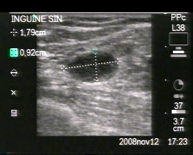
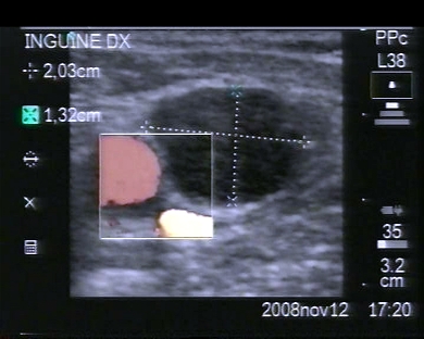
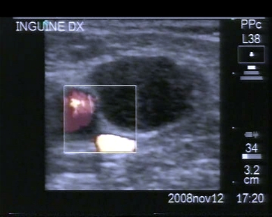
Inclusion date: 11 /02/2009Ultrasound made on: 12/11/2008 Instrument: Sonosite Patient's age: F 57 years old The ultrasound examination is carried out as a follow-up to a non Hodgkin’s lymphoma. The images and the video show lympho-adenomegalies, which are lumbo-aortic and located in the neck and in the inguinal area. In collaboration with: Dr. Pietro Vitali (oncologist–surgeon) - Jesi (AN) Presentation: Dr. Massimo Dolciotti - Ancona Digital processing: Andrea Dini - Ancona English translation: Prof.ssa Federica Sturani - England
|
|
|
|
|
|
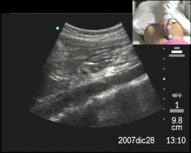
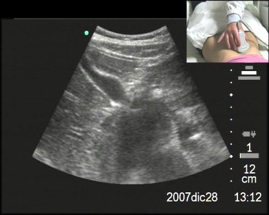
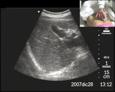
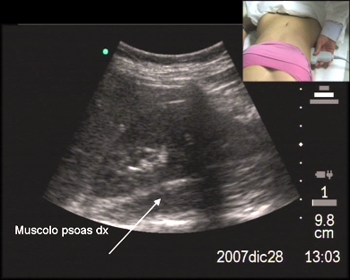
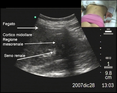
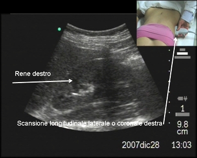
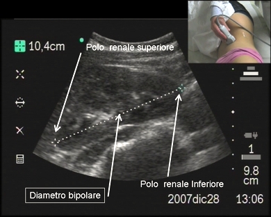
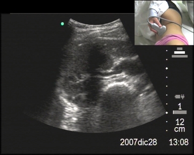
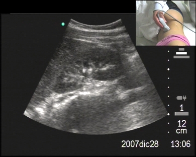
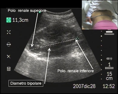
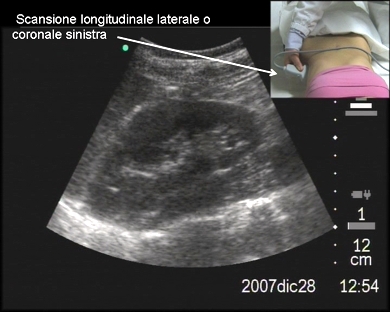
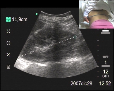
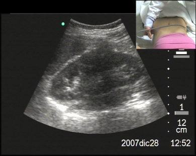
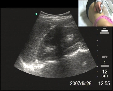
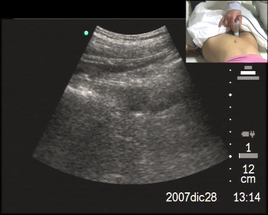
Inclusion date: 09 /02/2009Ultrasound made on: 28/12/2007 Instrument: Sonosite Patient's age: F 18 years old The ultrasound examination is carried out to study the right kidney and the suprarenal cavity. The images and the video show the right kidney with a regular echo-structure and morpho-volumetry. In addition to the supine position, the patient is examined while lying on the left side. Presentation: Dr. Massimo Dolciotti - Ancona Digital processing: Andrea Dini - Ancona English translation: Prof.ssa Federica Sturani - England
|
|
|
|
|
|
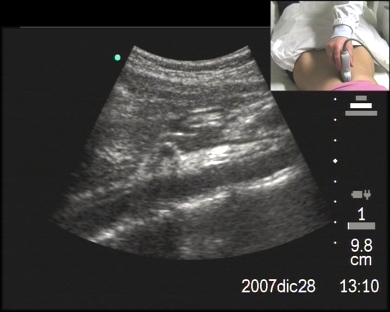
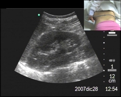
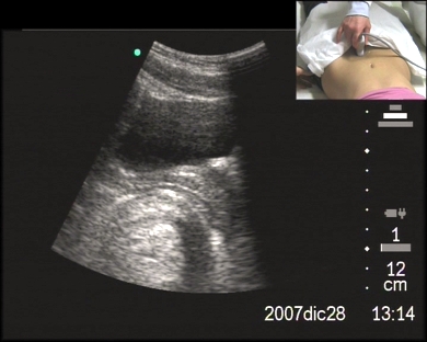
Inclusion date: 09 /02/2009Ultrasound made on: 28/12/2007 Instrument: Sonosite Patient's age: F 18 years old The ultrasound examination is carried out to study the left kidney and the suprarenal cavity. The images and the video show the left kidney with a regular echo-structure and morpho-volumetry. In addition to the supine position, the patient is examined while lying on the right side. Presentation: Dr. Massimo Dolciotti - Ancona Digital processing: Andrea Dini - Ancona English translation: Prof.ssa Federica Sturani - England
|
|
|
|
|
|
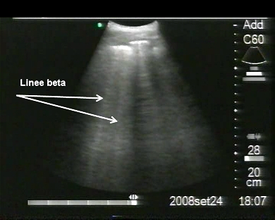
Inclusion date: 30 /01/2009Ultrasound made on: 24/09/2008 Instrument: Sonosite Patient's age: M 87 years old The ultrasound examination is carried out following cough and exertion dyspnea. The images and the videos show a small pleuric effusion to the right costophrenic angle. At the level of both the right and left middle-basal areas, the examination reveals beta lines which can be linked to a wet lung. In collaboration with: Dr. Pietro Vitali - Jesi (AN) Presentation: Dr. Massimo Dolciotti - Ancona Digital processing: Andrea Dini - Ancona English translation: Prof.ssa Federica Sturani - England
|
|
|
|
|
|
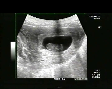
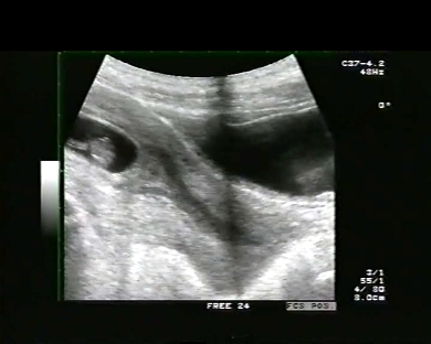
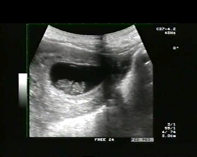
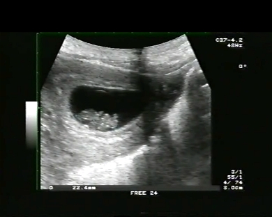
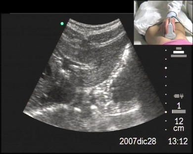
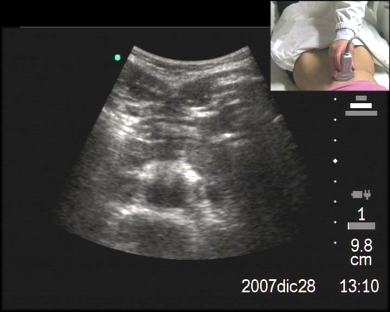
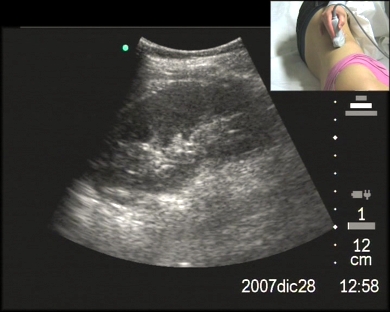
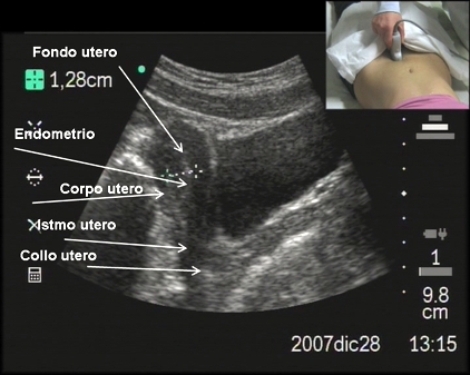
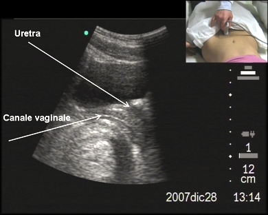
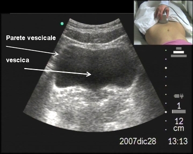
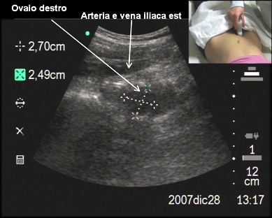
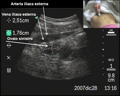
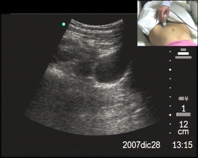
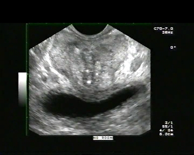
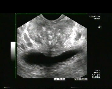
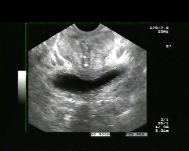
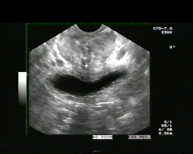
Inclusion date: 30 /01/2009Ultrasound made on: 28/12/2007 Instrument: Sonosite Patient's age: F 18 years old The ultrasound examination is carried out to study the female pelvis from a supra-pubic angle. The images and the video show the bladder, which is modestly full, and has got regular walls. The anteverted uterus is divided in its parts: neck, isthmus, body and fundus. The endometrium is in an excreting phase and is 12mm thick. The vaginal canal and the urethra are visible, and the right and left ovaries are highlighted. The ovaries have a regular echostructure and morphovolumetry. Presentation: Dr. Massimo Dolciotti - Ancona Digital processing: Andrea Dini - Ancona English translation: Prof.ssa Federica Sturani - England
|
|
|
|
|
|
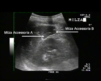
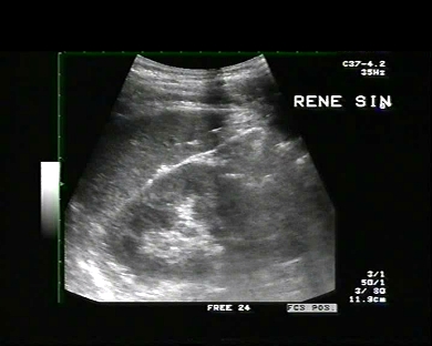
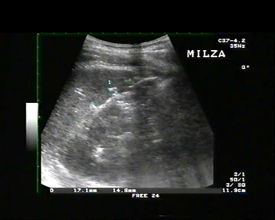
Inclusion date: 28 /01/2009Ultrasound made on: 02/01/2008 Instrument: Toshiba 380A Patient's age: M 32 years old The ultrasound examination is carried out following pains in the right hypochondrium. The images and the video show the spleen with a regular echo-morphovolumetry. In the splenic hilum an in the inferior pole there is evidence of oval and isoechoic images, which have a maximum diameter of 17mm and 14mm. They can be explained as two accessory spleens. Presentation: Dr. Massimo Dolciotti - Ancona Digital processing: Andrea Dini - Ancona English translation: Prof.ssa Federica Sturani - England
|
|
|
|
|
|
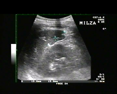
Inclusion date: 26 /01/2009Ultrasound made on: 25/01/2009 Instrument: Toshiba 380A Patient's age: F 66 years old The ultrasound examination is carried out following lumbar pains. The images and the video show the spleen with a regular echo-morphovolumetry. In the splenic hilum there is evidence of a isoechoic and oval image, 18x14mm in size, which can be explained as an accessory spleen. Presentation: Dr. Massimo Dolciotti - Ancona Digital processing: Andrea Dini - Ancona English translation: Prof.ssa Federica Sturani - England
|
|
|
|
|
|
Inclusion date: 26 /01/2009Ultrasound made on: 23/06/2008 Instrument: Toshiba 380A Patient's age: M 48 years old The ultrasound examination is carried out following dysuria, strangury , pollakiuria and scrotal pains located in the right epididymis. The images and video show the prostate with an echo-morphovolumetry above the norm, widespread intraglandular calcifications, enlarged right seminal vesicle and the right epididymis with increased volume. The ematochemical examinations show: PCR 12.9mg/dL, PSA 7.28 ng/dl, ESR 21mm/h, WBC 4,530, Lymphocyte 37%, Neutrophils 51%. Presentation: Dr. Massimo Dolciotti - Ancona Digital processing: Andrea Dini - Ancona English translation: Prof.ssa Federica Sturani - England
|
|
|
|
|
|
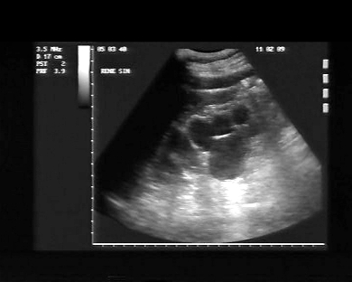
Inclusion date: 21 /01/2009Ultrasound made on: 27/10/2008 Instrument: Toshiba 380A Patient's age: M 52 years old The ultrasound examination is carried out following abdominal pains. The images and the video show the right kidney in an ectopic position in the right iliac fossa, in line with the bladder. The kidney shows a ureteral duplication. The left kidney is in regular position and has a regular morphovolumetry. The prostate has regular echo-structure and morphovolumetry. Presentation: Dr. Massimo Dolciotti - Ancona Digital processing: Andrea Dini - Ancona English translation: Prof.ssa Federica Sturani - England
|
|
|
|
|
|
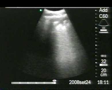
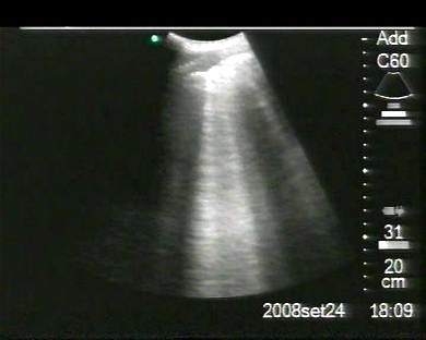
Inclusion date: 19 /01/2009Ultrasound made on: 12/01/2009 Instrument: Toshiba 380A Patient's age: M 67 years old VIDEOQUIZ: Which anomalies and pathologies of the urinary system does the patient under examination carry? For the discussion, we invite you to join the blog on the ecomiei website.Presentation: Dr. Massimo Dolciotti - Ancona Digital processing: Andrea Dini - Ancona English translation: Prof.ssa Federica Sturani - England
|
|
|
|
|
|
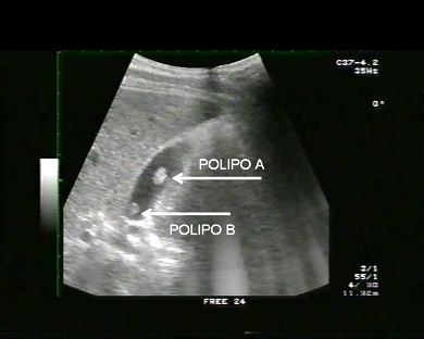
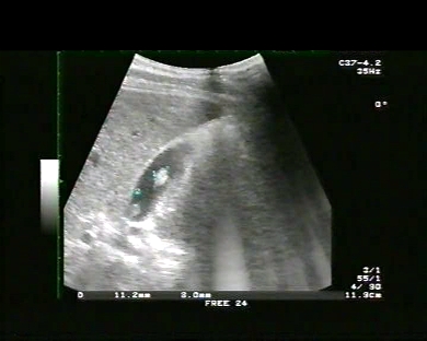
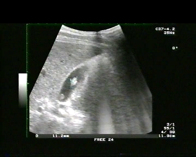
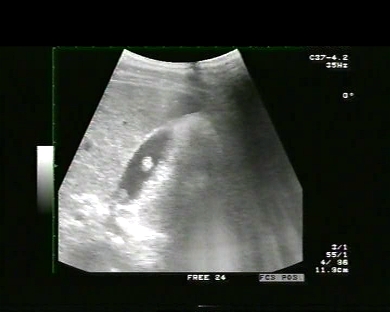
Inclusion date: 16 /01/2009Ultrasound made on: 01/02/2008 Instrument: Toshiba 380A Patient's age: F 34 years old The ultrasound examination is carried out following epigastric pains. The images and the video show two non-mobile isoechoic formations inside the cholecyst. The biggest of these formations measures 11mm of diameter and is located on the left lateral wall of the body; the second formation measures 8mm and is located on the right lateral wall, but deeper down the infundibulum. Both formations can be explained as polyps of the cholecyst. After a surgical evaluation, the patient underwent a cholecystectomy. In collaboration with: Dr. Quintilio Tomassetti - Ancona Presentation: Dr. Massimo Dolciotti - Ancona Digital processing: Andrea Dini - Ancona English translation: Prof.ssa Federica Sturani - England
|
|
|
|
|
|
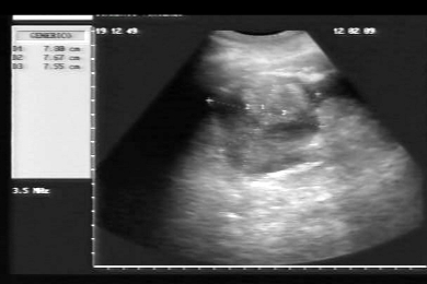
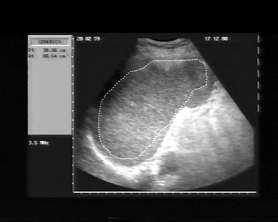
Inclusion date: 12 /01/2009Ultrasound made on: 17/12/2008 Instrument: Esaote Megas Patient's age: F 49 years old The ultrasound examination is carried out following pain on the left hypocondrium. The images and the video show the splenomegaly: the spleen has an echo-morphovolumetry above the norm, a bipolar diameter of 162mm and a section area bigger than 88 square mm. The examination also show an oval hyperechoic image on the superior pole of the spleen, which can be explained as a splenic angioma. In collaboration with: Dr. Daniele Lenti - Jesi (AN) Presentation: Dr. Massimo Dolciotti - Ancona Digital processing: Andrea Dini - Ancona English translation: Prof.ssa Federica Sturani - England
|
|
|
|
|
|
Inclusion date: 09 /01/2009Ultrasound made on: 22/09/2008 Instrument: Toshiba 380A Patient's age: F 47 years old The ultrasound examination is carried out following abdominal pains. The images and the video show an oval formation, which appears isoechoic against the cortex-medulla of the left kidney, on the left hypocondrium of a patient who had already undergone a splenectomy following a previous abdominal trauma with a spleen rupture. The formation can be explained as an accessory spleen. In addition, a hyperechoic image measuring 11x10 mm is visible in the inferior pole of the left kidney; this can be explained as a renal angiomyolipoma. Presentation: Dr. Massimo Dolciotti - Ancona Digital processing: Andrea Dini - Ancona English translation: Prof.ssa Federica Sturani - England
|
|
|
|
|
|
Inclusion date: 07 /01/2009Ultrasound made on: 12/12/2008 Instrument: Toshiba 380A Patient's age: M 14 years old The ultrasound examination is carried out following abdominal pains. The images and the video show in the left hypocondrium the spleen with a normal echo-morphovolumetry. In the splenic hilum there is evidence of an oval, isoechoic image measuring 19x20mm, which can be explained as an accessory spleen. Presentation: Dr. Massimo Dolciotti - Ancona Digital processing: Andrea Dini - Ancona English translation: Prof.ssa Federica Sturani - England
|
|
|
|
|
|
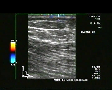
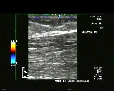
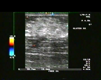
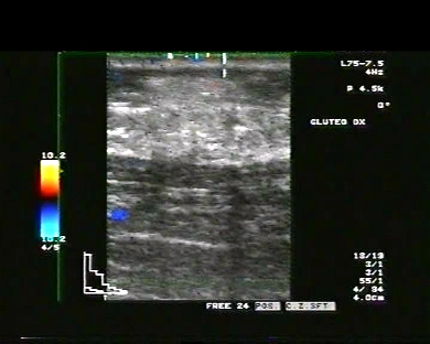
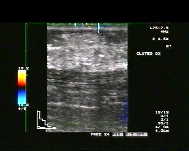
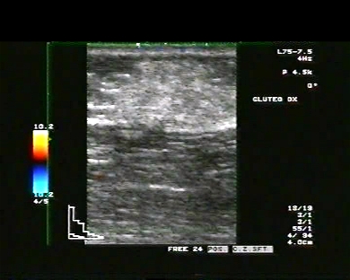
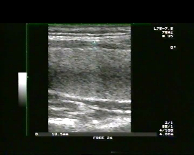
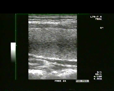
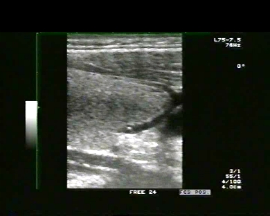
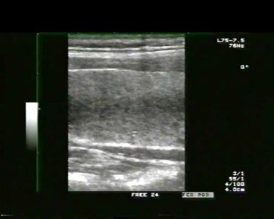
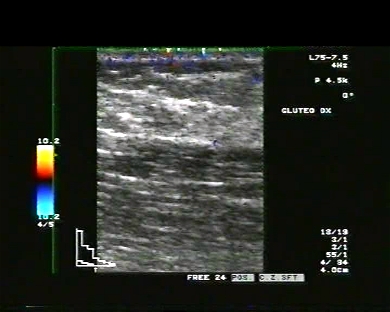
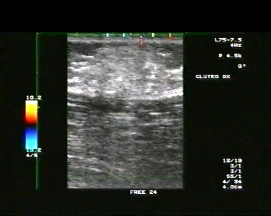
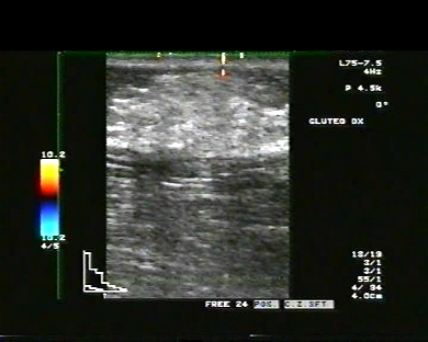
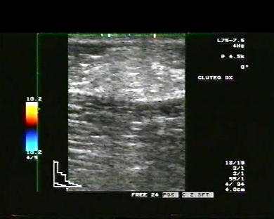
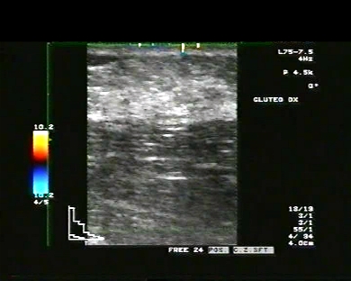
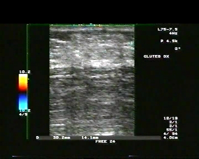
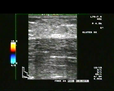
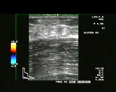
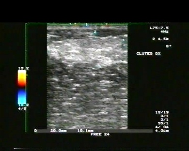
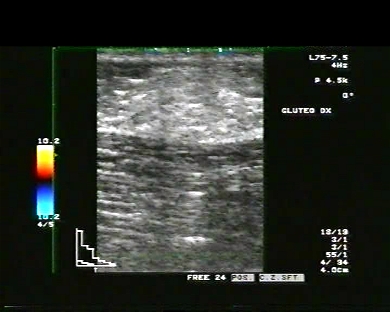
Inclusion date: 05 /01/2009Ultrasound made on: 29/12/2008 Instrument: Toshiba 380A Patient's age: F 38 years old The ultrasound examination is carried out following a painful swelling in the external higher region of the right gluteus, which started a few days earlier and two weeks after an intramuscular injection of analgesic. The images and the video show an oval formation, expanding from the subcutaneous tissue to the muscle, and being 14mm deep and 30mm wide. This can be explained as a muscular infiltrate. Presentation: Dr. Massimo Dolciotti - Ancona Digital processing: Andrea Dini - Ancona English translation: Prof.ssa Federica Sturani - England
|
|
|
|
|
|
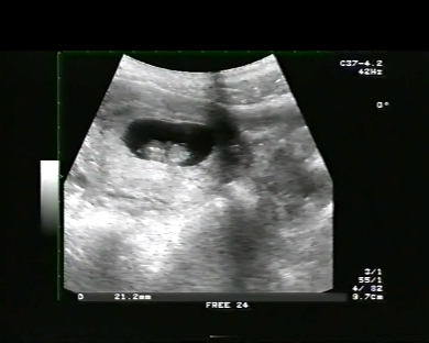
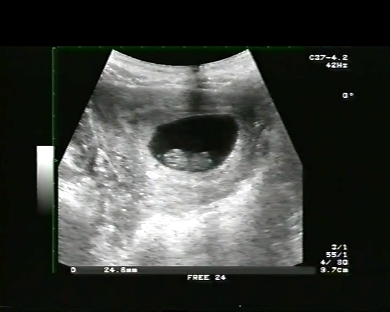
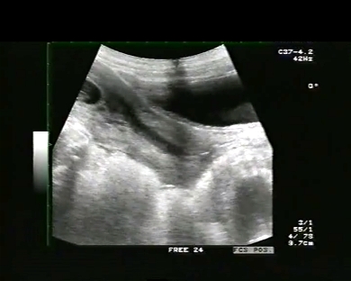
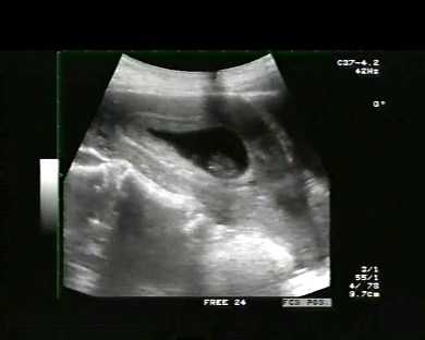
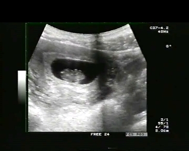
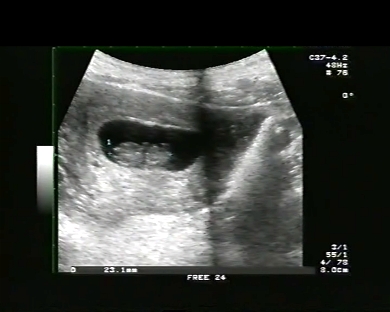
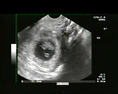
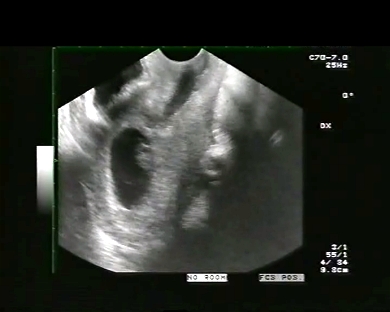
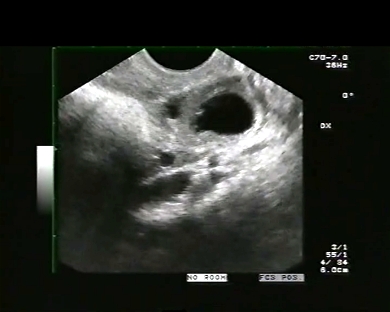
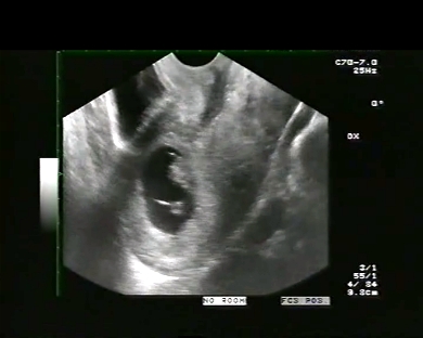
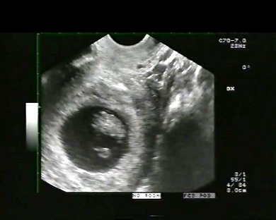
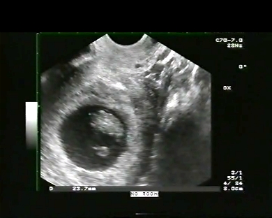
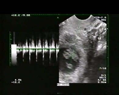
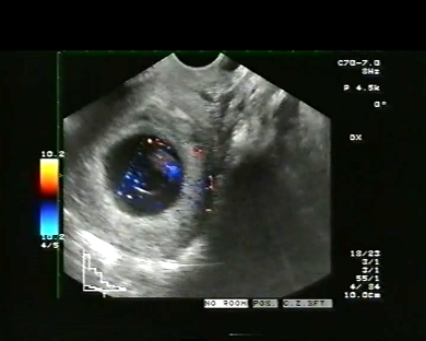
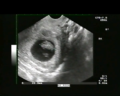
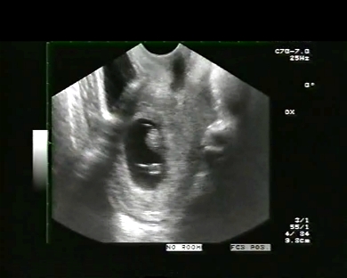
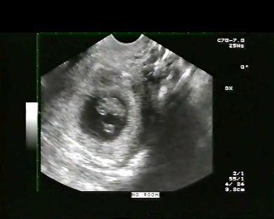
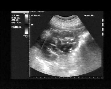
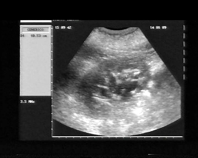
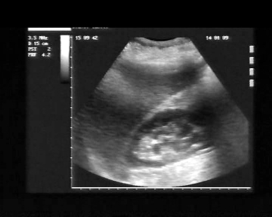
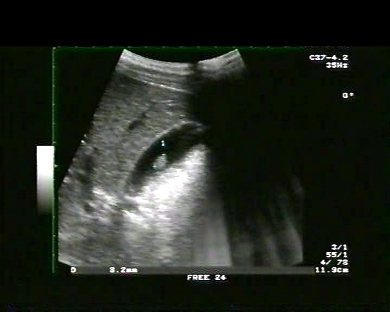
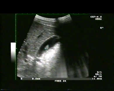
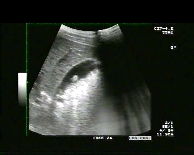
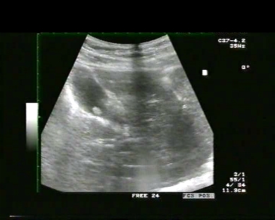
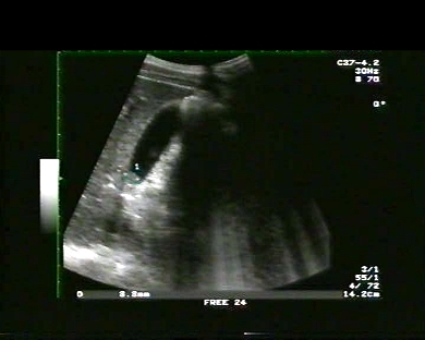
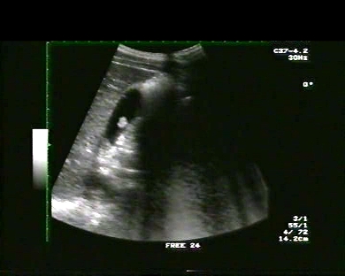
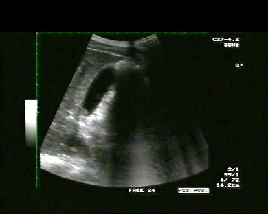
Inclusion date: 05 /01/2009Ultrasound made on: 12/08/2008 Instrument: Toshiba 380A Patient's age: F 28 years old The ultrasound examination of the pelvis in a patient in amenorrhea for 8 weeks is carried out from a suprapubic and endo-cavitary position. The images and the video show the pregnancy state, with the presence of foetal movements and heartbeat, a Crown-rump Length diameter of 21.2 mm and corpus luteum in the right ovary. Presentation: Dr. Massimo Dolciotti - Ancona Digital processing: Andrea Dini - Ancona English translation: Prof.ssa Federica Sturani - England
|
|
|
|
|
|
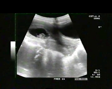
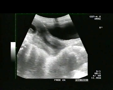
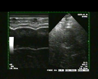
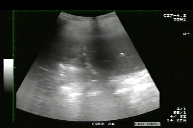
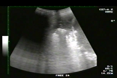
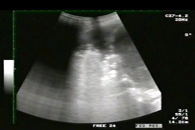
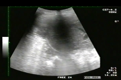
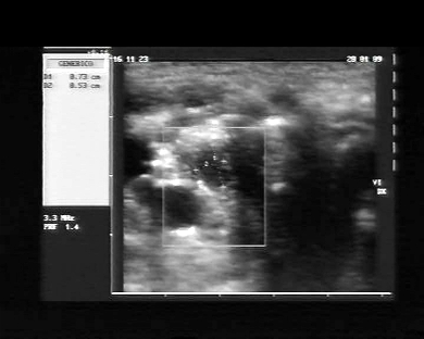
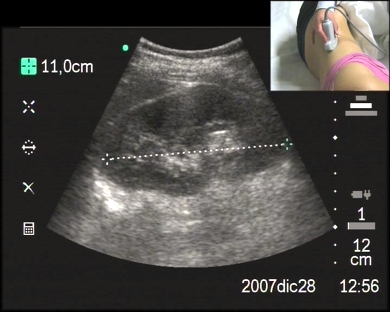
Inclusion date: 02 /01/2009Ultrasound made on: 14/11/2008 Instrument: Toshiba 380A Patient's age: F 87 years old The ultrasound examination of the chest and the abdomen is carried out following recurring lipothymias and dyspnea caused by minimal efforts. The images and the video show an aneurism to the abdominal aorta positioned underneath the kidney, with a maximum diameter shown by the ultrasound as being 77mm and lately estimated by the abdomen CAT to be 7.5 cm with a caudal extension to the “Carrefour”, with a true lumen of 4.5 cm and parietal calcifications. Presentation: Dr. Massimo Dolciotti - Ancona Digital processing: Andrea Dini - Ancona English translation: Prof.ssa Federica Sturani - England
|
|
|
|


 ù
ù